4OB2

Crystal Structure of Nitrile Hydratase from Pseudonocardia thermophila bound to Butaneboronic Acid via Crystal Soaking


Experimental Data Snapshot

  • Method: X-RAY DIFFRACTION
  • Resolution: 1.52 Å
  • R-Value Free: 0.183 
  • R-Value Work: 0.152 
  • R-Value Observed: 0.153 

wwPDB Validation   3D Report Full Report


This is version 1.1 of the entry. See complete history


Literature

The active site sulfenic acid ligand in nitrile hydratases can function as a nucleophile.

Martinez, S.Wu, R.Sanishvili, R.Liu, D.Holz, R.

(2014) J Am Chem Soc 136: 1186-1189

  • DOI: https://doi.org/10.1021/ja410462j
  • Primary Citation of Related Structures:  
    4OB0, 4OB1, 4OB2, 4OB3

  • PubMed Abstract: 

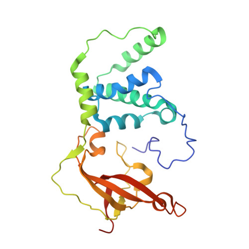
    Nitrile hydratase (NHase) catalyzes the hydration of nitriles to their corresponding commercially valuable amides at ambient temperatures and physiological pH. Several reaction mechanisms have been proposed for NHase enzymes; however, the source of the nucleophile remains a mystery. Boronic acids have been shown to be potent inhibitors of numerous hydrolytic enzymes due to the open shell of boron, which allows it to expand from a trigonal planar (sp(2)) form to a tetrahedral form (sp(3)). Therefore, we examined the inhibition of the Co-type NHase from Pseudonocardia thermophila JCM 3095 (PtNHase) by boronic acids via kinetics and X-ray crystallography. Both 1-butaneboronic acid (BuBA) and phenylboronic acid (PBA) function as potent competitive inhibitors of PtNHase. X-ray crystal structures for BuBA and PBA complexed to PtNHase were solved and refined at 1.5, 1.6, and 1.2 Å resolution. The resulting PtNHase-boronic acid complexes represent a "snapshot" of reaction intermediates and implicate the cysteine-sulfenic acid ligand as the catalytic nucleophile, a heretofore unknown role for the αCys(113)-OH sulfenic acid ligand. Based on these data, a new mechanism of action for the hydration of nitriles by NHase is presented.


  • Organizational Affiliation

    Department of Chemistry, Marquette University , Milwaukee, Wisconsin 53201, United States.


Macromolecules
Find similar proteins by:  (by identity cutoff)  |  3D Structure
Entity ID: 1
MoleculeChains Sequence LengthOrganismDetailsImage
Cobalt-containing nitrile hydratase subunit alpha209Pseudonocardia thermophilaMutation(s): 0 
EC: 4.2.1.84
UniProt
Find proteins for Q7SID2 (Pseudonocardia thermophila)
Explore Q7SID2 
Go to UniProtKB:  Q7SID2
Entity Groups  
Sequence Clusters30% Identity50% Identity70% Identity90% Identity95% Identity100% Identity
UniProt GroupQ7SID2
Sequence Annotations
Expand
  • Reference Sequence
Find similar proteins by:  (by identity cutoff)  |  3D Structure
Entity ID: 2
MoleculeChains Sequence LengthOrganismDetailsImage
Cobalt-containing nitrile hydratase subunit beta233Pseudonocardia thermophilaMutation(s): 0 
EC: 4.2.1.84
UniProt
Find proteins for Q7SID3 (Pseudonocardia thermophila)
Explore Q7SID3 
Go to UniProtKB:  Q7SID3
Entity Groups  
Sequence Clusters30% Identity50% Identity70% Identity90% Identity95% Identity100% Identity
UniProt GroupQ7SID3
Sequence Annotations
Expand
  • Reference Sequence
Small Molecules
Ligands 4 Unique
IDChains Name / Formula / InChI Key2D Diagram3D Interactions
BUB
Query on BUB

Download Ideal Coordinates CCD File 
C [auth A]1-BUTANE BORONIC ACID
C4 H11 B O2
QPKFVRWIISEVCW-UHFFFAOYSA-N
GOL
Query on GOL

Download Ideal Coordinates CCD File 
E [auth A]
F [auth A]
G [auth A]
H [auth A]
R [auth B]
E [auth A],
F [auth A],
G [auth A],
H [auth A],
R [auth B],
S [auth B],
T [auth B],
U [auth B]
GLYCEROL
C3 H8 O3
PEDCQBHIVMGVHV-UHFFFAOYSA-N
CO
Query on CO

Download Ideal Coordinates CCD File 
D [auth A]COBALT (II) ION
Co
XLJKHNWPARRRJB-UHFFFAOYSA-N
MOH
Query on MOH

Download Ideal Coordinates CCD File 
AA [auth B]
BA [auth B]
CA [auth B]
DA [auth B]
EA [auth B]
AA [auth B],
BA [auth B],
CA [auth B],
DA [auth B],
EA [auth B],
FA [auth B],
GA [auth B],
HA [auth B],
I [auth A],
IA [auth B],
J [auth A],
JA [auth B],
K [auth A],
L [auth A],
M [auth A],
N [auth A],
O [auth A],
P [auth A],
Q [auth A],
V [auth B],
W [auth B],
X [auth B],
Y [auth B],
Z [auth B]
METHANOL
C H4 O
OKKJLVBELUTLKV-UHFFFAOYSA-N
Modified Residues  1 Unique
IDChains TypeFormula2D DiagramParent
CSD
Query on CSD
A
L-PEPTIDE LINKINGC3 H7 N O4 SCYS
Experimental Data & Validation

Experimental Data

  • Method: X-RAY DIFFRACTION
  • Resolution: 1.52 Å
  • R-Value Free: 0.183 
  • R-Value Work: 0.152 
  • R-Value Observed: 0.153 
  • Space Group: P 32 2 1
Unit Cell:
Length ( Å )Angle ( ˚ )
a = 65.601α = 90
b = 65.601β = 90
c = 185.658γ = 120
Software Package:
Software NamePurpose
HKL-2000data collection
PHASESphasing
PHENIXrefinement
HKL-2000data reduction
HKL-2000data scaling

Structure Validation

View Full Validation Report


Entry History 

Deposition Data

Revision History  (Full details and data files)

  • Version 1.0: 2014-11-26
    Type: Initial release
  • Version 1.1: 2019-11-20
    Changes: Database references, Derived calculations