


SAICAR synthetase (Type-1) in complex with GMP
Manjunath, K., Jeyakanthan, J., Sekar, K.To be published.
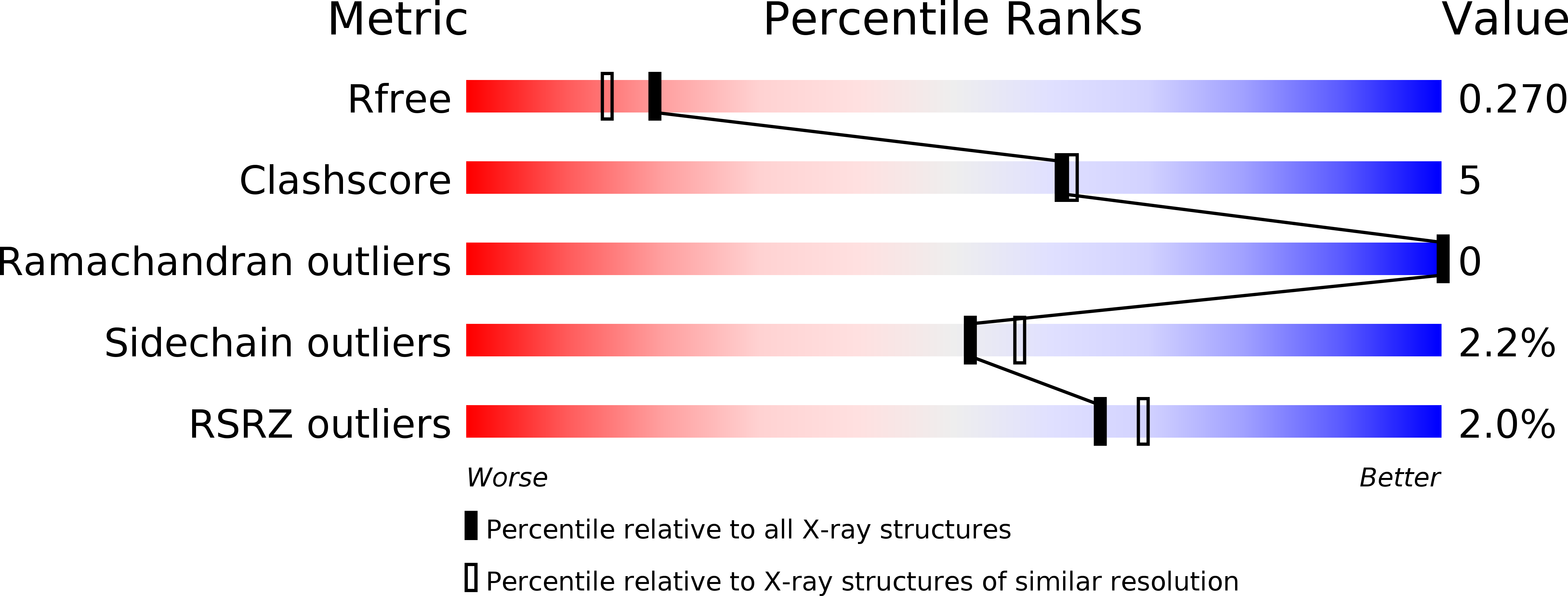
Experimental Data Snapshot
Entity ID: 1 | |||||
|---|---|---|---|---|---|
| Molecule | Chains | Sequence Length | Organism | Details | Image |
| Phosphoribosylaminoimidazole-succinocarboxamide synthase | 238 | Pyrococcus horikoshii OT3 | Mutation(s): 0 Gene Names: PH0239, purC EC: 6.3.2.6 |  | |
UniProt | |||||
Find proteins for O57978 (Pyrococcus horikoshii (strain ATCC 700860 / DSM 12428 / JCM 9974 / NBRC 100139 / OT-3)) Explore O57978 Go to UniProtKB: O57978 | |||||
Entity Groups | |||||
| Sequence Clusters | 30% Identity50% Identity70% Identity90% Identity95% Identity100% Identity | ||||
| UniProt Group | O57978 | ||||
Sequence AnnotationsExpand | |||||
| |||||
| Ligands 4 Unique | |||||
|---|---|---|---|---|---|
| ID | Chains | Name / Formula / InChI Key | 2D Diagram | 3D Interactions | |
| 5GP Query on 5GP | J [auth A], T [auth B] | GUANOSINE-5'-MONOPHOSPHATE C10 H14 N5 O8 P RQFCJASXJCIDSX-UUOKFMHZSA-N |  | ||
| CD Query on CD | C [auth A] D [auth A] E [auth A] F [auth A] G [auth A] | CADMIUM ION Cd WLZRMCYVCSSEQC-UHFFFAOYSA-N |  | ||
| PO4 Query on PO4 | K [auth A], U [auth B] | PHOSPHATE ION O4 P NBIIXXVUZAFLBC-UHFFFAOYSA-K |  | ||
| BU1 Query on BU1 | L [auth A], V [auth B] | 1,4-BUTANEDIOL C4 H10 O2 WERYXYBDKMZEQL-UHFFFAOYSA-N |  | ||
| Modified Residues 1 Unique | |||||
|---|---|---|---|---|---|
| ID | Chains | Type | Formula | 2D Diagram | Parent |
| MSE Query on MSE | A, B | L-PEPTIDE LINKING | C5 H11 N O2 Se |  | MET |
| Length ( Å ) | Angle ( ˚ ) |
|---|---|
| a = 94.81 | α = 90 |
| b = 94.81 | β = 90 |
| c = 148.051 | γ = 120 |
| Software Name | Purpose |
|---|---|
| MOSFLM | data reduction |
| SCALA | data scaling |
| PHASER | phasing |
| REFMAC | refinement |
| PDB_EXTRACT | data extraction |
| MAR345dtb | data collection |
RCSB PDB (citation) is hosted by
RCSB PDB is a member of the

