4NN9

REFINED ATOMIC STRUCTURES OF N9 SUBTYPE INFLUENZA VIRUS NEURAMINIDASE AND ESCAPE MUTANTS


Experimental Data Snapshot

  • Method: X-RAY DIFFRACTION
  • Resolution: 2.30 Å
  • R-Value Work: 0.163 

wwPDB Validation   3D Report Full Report


Ligand Structure Quality Assessment 


This is version 2.0 of the entry. See complete history


Literature

Refined atomic structures of N9 subtype influenza virus neuraminidase and escape mutants.

Tulip, W.R.Varghese, J.N.Baker, A.T.van Donkelaar, A.Laver, W.G.Webster, R.G.Colman, P.M.

(1991) J Mol Biol 221: 487-497

  • DOI: https://doi.org/10.1016/0022-2836(91)80069-7
  • Primary Citation of Related Structures:  
    3NN9, 4NN9, 5NN9, 6NN9

  • PubMed Abstract: 

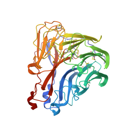
    The crystal structure of the N9 subtype neuraminidase of influenza virus was refined by simulated annealing and conventional techniques to an R-factor of 0.172 for data in the resolution range 6.0 to 2.2 A. The r.m.s. deviation from ideal values of bond lengths is 0.014 A. The structure is similar to that of N2 subtype neuraminidase both in secondary structure elements and in their connections. The three-dimensional structures of several escape mutants of neuraminidase, selected with antineuraminidase monoclonal antibodies, are also reported. In every case, structural changes associated with the point mutation are confined to the mutation site or to residues that are spatially immediately adjacent to it. The failure of antisera to cross-react between N2 and N9 subtypes may be correlated with the absence of conserved, contiguous surface structures of area 700 A2 or more.


  • Organizational Affiliation

    CSIRO Division of Biomolecular Engineering, Parkville, Australia.


Macromolecules
Find similar proteins by:  (by identity cutoff)  |  3D Structure
Entity ID: 1
MoleculeChains Sequence LengthOrganismDetailsImage
NEURAMINIDASE N9388Influenza A virus (A/tern/Australia/G70C/1975(H11N9))Mutation(s): 0 
EC: 3.2.1.18
UniProt
Find proteins for P03472 (Influenza A virus (strain A/Tern/Australia/G70C/1975 H11N9))
Explore P03472 
Go to UniProtKB:  P03472
Entity Groups  
Sequence Clusters30% Identity50% Identity70% Identity90% Identity95% Identity100% Identity
UniProt GroupP03472
Sequence Annotations
Expand
  • Reference Sequence
Oligosaccharides

Help

Entity ID: 2
MoleculeChains Length2D Diagram Glycosylation3D Interactions
alpha-D-mannopyranose-(1-2)-alpha-D-mannopyranose-(1-2)-alpha-D-mannopyranose-(1-3)-[alpha-D-mannopyranose-(1-6)]alpha-D-mannopyranose-(1-4)-2-acetamido-2-deoxy-beta-D-glucopyranose-(1-4)-2-acetamido-2-deoxy-beta-D-glucopyranose
B
7N-Glycosylation
Glycosylation Resources
GlyTouCan:  G57082ZE
GlyCosmos:  G57082ZE
GlyGen:  G57082ZE
Experimental Data & Validation

Experimental Data

  • Method: X-RAY DIFFRACTION
  • Resolution: 2.30 Å
  • R-Value Work: 0.163 
  • Space Group: I 4 3 2
Unit Cell:
Length ( Å )Angle ( ˚ )
a = 185.1α = 90
b = 185.1β = 90
c = 185.1γ = 90
Software Package:
Software NamePurpose
X-PLORrefinement

Structure Validation

View Full Validation Report


Ligand Structure Quality Assessment 


Entry History 

Deposition Data

Revision History  (Full details and data files)

  • Version 1.0: 1992-07-15
    Type: Initial release
  • Version 1.1: 2008-03-25
    Changes: Version format compliance
  • Version 1.2: 2011-07-13
    Changes: Source and taxonomy, Version format compliance
  • Version 2.0: 2020-07-29
    Type: Remediation
    Reason: Carbohydrate remediation
    Changes: Advisory, Atomic model, Data collection, Database references, Derived calculations, Other, Structure summary