4NL8

PriA Helicase Bound to SSB C-terminal Tail Peptide


Experimental Data Snapshot

  • Method: X-RAY DIFFRACTION
  • Resolution: 4.08 Å
  • R-Value Free: 0.298 
  • R-Value Work: 0.240 
  • R-Value Observed: 0.243 

wwPDB Validation   3D Report Full Report


This is version 1.2 of the entry. See complete history


Literature

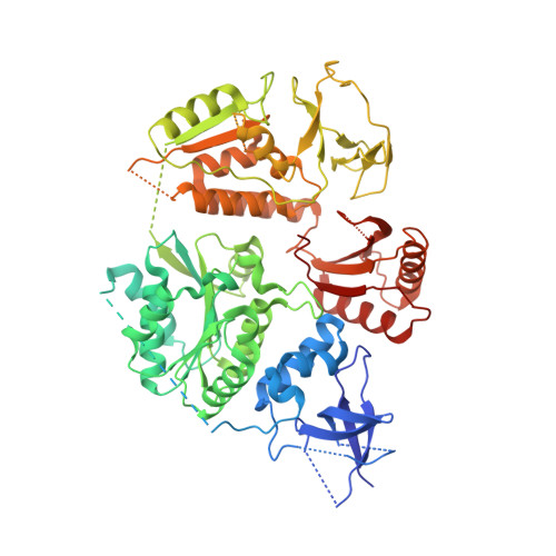
Structural mechanisms of PriA-mediated DNA replication restart.

Bhattacharyya, B.George, N.P.Thurmes, T.M.Zhou, R.Jani, N.Wessel, S.R.Sandler, S.J.Ha, T.Keck, J.L.

(2014) Proc Natl Acad Sci U S A 111: 1373-1378

  • DOI: https://doi.org/10.1073/pnas.1318001111
  • Primary Citation of Related Structures:  
    4NL4, 4NL8

  • PubMed Abstract: 

    Collisions between cellular DNA replication machinery (replisomes) and damaged DNA or immovable protein complexes can dissociate replisomes before the completion of replication. This potentially lethal problem is resolved by cellular "replication restart" reactions that recognize the structures of prematurely abandoned replication forks and mediate replisomal reloading. In bacteria, this essential activity is orchestrated by the PriA DNA helicase, which identifies replication forks via structure-specific DNA binding and interactions with fork-associated ssDNA-binding proteins (SSBs). However, the mechanisms by which PriA binds replication fork DNA and coordinates subsequent replication restart reactions have remained unclear due to the dearth of high-resolution structural information available for the protein. Here, we describe the crystal structures of full-length PriA and PriA bound to SSB. The structures reveal a modular arrangement for PriA in which several DNA-binding domains surround its helicase core in a manner that appears to be poised for binding to branched replication fork DNA structures while simultaneously allowing complex formation with SSB. PriA interaction with SSB is shown to modulate SSB/DNA complexes in a manner that exposes a potential replication initiation site. From these observations, a model emerges to explain how PriA links recognition of diverse replication forks to replication restart.


  • Organizational Affiliation

    Department of Biomolecular Chemistry, School of Medicine and Public Health, University of Wisconsin, Madison, WI 53706.


Macromolecules

Find similar proteins by:  Sequence   |   3D Structure  

Entity ID: 1
MoleculeChains Sequence LengthOrganismDetailsImage
Single-stranded DNA-binding proteinA [auth C],
C [auth D],
E [auth F]
10Klebsiella pneumoniae subsp. pneumoniae HS11286Mutation(s): 0 
Gene Names: KPHS_02830ssb c-terminal peptide
UniProt
Find proteins for A0A0H3GL04 (Klebsiella pneumoniae subsp. pneumoniae (strain HS11286))
Explore A0A0H3GL04 
Go to UniProtKB:  A0A0H3GL04
Entity Groups  
Sequence Clusters30% Identity50% Identity70% Identity90% Identity95% Identity100% Identity
UniProt GroupA0A0H3GL04
Sequence Annotations
Expand
  • Reference Sequence
Find similar proteins by:  (by identity cutoff)  |  3D Structure
Entity ID: 2
MoleculeChains Sequence LengthOrganismDetailsImage
Primosome assembly protein PriAB [auth A],
D [auth B],
F [auth E]
747Klebsiella pneumoniae subsp. pneumoniae MGH 78578Mutation(s): 0 
Gene Names: KPN78578_41850KPN_04230priA
UniProt
Find proteins for A6TGC5 (Klebsiella pneumoniae subsp. pneumoniae (strain ATCC 700721 / MGH 78578))
Explore A6TGC5 
Go to UniProtKB:  A6TGC5
Entity Groups  
Sequence Clusters30% Identity50% Identity70% Identity90% Identity95% Identity100% Identity
UniProt GroupA6TGC5
Sequence Annotations
Expand
  • Reference Sequence
Experimental Data & Validation

Experimental Data

  • Method: X-RAY DIFFRACTION
  • Resolution: 4.08 Å
  • R-Value Free: 0.298 
  • R-Value Work: 0.240 
  • R-Value Observed: 0.243 
  • Space Group: P 21 21 21
Unit Cell:
Length ( Å )Angle ( ˚ )
a = 106.61α = 90
b = 153.248β = 90
c = 192.373γ = 90
Software Package:
Software NamePurpose
ADSCdata collection
PHASERphasing
REFMACrefinement
HKL-2000data reduction
HKL-2000data scaling

Structure Validation

View Full Validation Report


Entry History 

Deposition Data

Revision History  (Full details and data files)

  • Version 1.0: 2014-01-08
    Type: Initial release
  • Version 1.1: 2014-02-12
    Changes: Database references
  • Version 1.2: 2024-02-28
    Changes: Data collection, Database references, Derived calculations, Refinement description