4MH1

Crystal structure and functional studies of quinoprotein L-sorbose dehydrogenase from Ketogulonicigenium vulgare Y25


Experimental Data Snapshot

  • Method: X-RAY DIFFRACTION
  • Resolution: 2.70 Å
  • R-Value Free: 0.232 
  • R-Value Work: 0.226 
  • R-Value Observed: 0.226 

wwPDB Validation   3D Report Full Report


Ligand Structure Quality Assessment 


This is version 1.1 of the entry. See complete history


Literature

Crystal structure of L-sorbose dehydrogenase, a pyrroloquinoline quinone-dependent enzyme with homodimeric assembly, from Ketogulonicigenium vulgare

Han, X.Xiong, X.Jiang, D.Chen, S.Huang, E.Zhang, W.Liu, X.

(2014) Biotechnol Lett 36: 1001-1008

  • DOI: https://doi.org/10.1007/s10529-013-1446-5
  • Primary Citation of Related Structures:  
    4MH1

  • PubMed Abstract: 

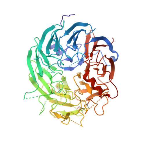
    The crystal structure of the L-sorbose dehydrogenase (SDH) from Ketogulonicigenium vulgare Y25 has been determined at 2.7 Å resolution using the molecular replacement method. The overall structure of SDH is similar to that of other quinoprotein dehydrogenases; consisting of an eight bladed β-propeller PQQ domain and protrusion loops. We identified a stable homodimer in crystal and demonstrated its existence in solution by sedimentation velocity measurement. By biochemical characterization of the SDH in vitro, using L-sorbose as substrate and cytochrome c551 as electron acceptor, we revealed cytochrome c551 acting as physiological primary electron acceptor for SDH.


  • Organizational Affiliation

    State Key Laboratory of Medicinal Chemical Biology, College of Life Sciences, Nankai University, Tianjin, 300071, China, hxdon@126.com.


Macromolecules
Find similar proteins by:  (by identity cutoff)  |  3D Structure
Entity ID: 1
MoleculeChains Sequence LengthOrganismDetailsImage
Sorbose dehydrogenase
A, B
560Ketogulonicigenium vulgare Y25Mutation(s): 0 
Entity Groups  
Sequence Clusters30% Identity50% Identity70% Identity90% Identity95% Identity100% Identity
Sequence Annotations
Expand
  • Reference Sequence
Small Molecules
Ligands 2 Unique
IDChains Name / Formula / InChI Key2D Diagram3D Interactions
PQQ
Query on PQQ

Download Ideal Coordinates CCD File 
C [auth B]PYRROLOQUINOLINE QUINONE
C14 H6 N2 O8
MMXZSJMASHPLLR-UHFFFAOYSA-N
CA
Query on CA

Download Ideal Coordinates CCD File 
D [auth B]CALCIUM ION
Ca
BHPQYMZQTOCNFJ-UHFFFAOYSA-N
Experimental Data & Validation

Experimental Data

  • Method: X-RAY DIFFRACTION
  • Resolution: 2.70 Å
  • R-Value Free: 0.232 
  • R-Value Work: 0.226 
  • R-Value Observed: 0.226 
  • Space Group: P 4 3 2
Unit Cell:
Length ( Å )Angle ( ˚ )
a = 201.53α = 90
b = 201.53β = 90
c = 201.53γ = 90
Software Package:
Software NamePurpose
HKL-2000data collection
BALBESphasing
PHENIXrefinement
HKL-2000data reduction
HKL-2000data scaling

Structure Validation

View Full Validation Report


Ligand Structure Quality Assessment 


Entry History 

Deposition Data

Revision History  (Full details and data files)

  • Version 1.0: 2014-07-23
    Type: Initial release
  • Version 1.1: 2023-11-08
    Changes: Data collection, Database references, Derived calculations, Refinement description