4MDP

Crystal structure of a GH1 beta-glucosidase from the fungus Humicola insolens in complex with glucose


Experimental Data Snapshot

  • Method: X-RAY DIFFRACTION
  • Resolution: 2.05 Å
  • R-Value Free: 0.214 
  • R-Value Work: 0.163 
  • R-Value Observed: 0.165 

wwPDB Validation   3D Report Full Report


Ligand Structure Quality Assessment 


This is version 1.3 of the entry. See complete history


Literature

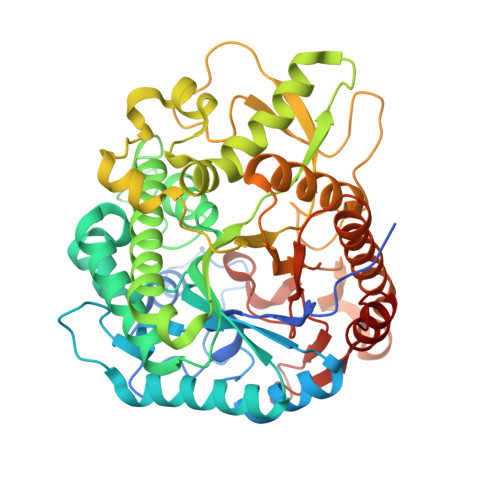
Structural basis for glucose tolerance in GH1 beta-glucosidases.

Giuseppe, P.O.Souza, T.A.C.B.Souza, F.H.M.Zanphorlin, L.M.Machado, C.B.Ward, R.J.Jorge, J.A.Furriel, R.P.M.Murakami, M.T.

(2014) Acta Crystallogr D Biol Crystallogr 70: 1631-1639

  • DOI: https://doi.org/10.1107/S1399004714006920
  • Primary Citation of Related Structures:  
    4MDO, 4MDP

  • PubMed Abstract: 

    Product inhibition of β-glucosidases (BGs) by glucose is considered to be a limiting step in enzymatic technologies for plant-biomass saccharification. Remarkably, some β-glucosidases belonging to the GH1 family exhibit unusual properties, being tolerant to, or even stimulated by, high glucose concentrations. However, the structural basis for the glucose tolerance and stimulation of BGs is still elusive. To address this issue, the first crystal structure of a fungal β-glucosidase stimulated by glucose was solved in native and glucose-complexed forms, revealing that the shape and electrostatic properties of the entrance to the active site, including the +2 subsite, determine glucose tolerance. The aromatic Trp168 and the aliphatic Leu173 are conserved in glucose-tolerant GH1 enzymes and contribute to relieving enzyme inhibition by imposing constraints at the +2 subsite that limit the access of glucose to the -1 subsite. The GH1 family β-glucosidases are tenfold to 1000-fold more glucose tolerant than GH3 BGs, and comparative structural analysis shows a clear correlation between active-site accessibility and glucose tolerance. The active site of GH1 BGs is located in a deep and narrow cavity, which is in contrast to the shallow pocket in the GH3 family BGs. These findings shed light on the molecular basis for glucose tolerance and indicate that GH1 BGs are more suitable than GH3 BGs for biotechnological applications involving plant cell-wall saccharification.


  • Organizational Affiliation

    Brazilian Biosciences National Laboratory, National Center for Research in Energy and Materials, CP 6192, 13083-970 Campinas-SP, Brazil.


Macromolecules
Find similar proteins by:  (by identity cutoff)  |  3D Structure
Entity ID: 1
MoleculeChains Sequence LengthOrganismDetailsImage
Beta-glucosidase499[Humicola] grisea var. thermoideaMutation(s): 0 
Gene Names: bgl4
UniProt
Find proteins for O93784 (Humicola grisea var. thermoidea)
Explore O93784 
Go to UniProtKB:  O93784
Entity Groups  
Sequence Clusters30% Identity50% Identity70% Identity90% Identity95% Identity100% Identity
UniProt GroupO93784
Sequence Annotations
Expand
  • Reference Sequence
Small Molecules
Ligands 4 Unique
IDChains Name / Formula / InChI Key2D Diagram3D Interactions
BGC
Query on BGC

Download Ideal Coordinates CCD File 
B [auth A]beta-D-glucopyranose
C6 H12 O6
WQZGKKKJIJFFOK-VFUOTHLCSA-N
PEG
Query on PEG

Download Ideal Coordinates CCD File 
C [auth A],
D [auth A],
E [auth A]
DI(HYDROXYETHYL)ETHER
C4 H10 O3
MTHSVFCYNBDYFN-UHFFFAOYSA-N
PO4
Query on PO4

Download Ideal Coordinates CCD File 
F [auth A],
G [auth A]
PHOSPHATE ION
O4 P
NBIIXXVUZAFLBC-UHFFFAOYSA-K
GOL
Query on GOL

Download Ideal Coordinates CCD File 
H [auth A]GLYCEROL
C3 H8 O3
PEDCQBHIVMGVHV-UHFFFAOYSA-N
Experimental Data & Validation

Experimental Data

  • Method: X-RAY DIFFRACTION
  • Resolution: 2.05 Å
  • R-Value Free: 0.214 
  • R-Value Work: 0.163 
  • R-Value Observed: 0.165 
  • Space Group: P 65 2 2
Unit Cell:
Length ( Å )Angle ( ˚ )
a = 113.567α = 90
b = 113.567β = 90
c = 178.74γ = 120
Software Package:
Software NamePurpose
SCALEPACKdata scaling
REFMACrefinement
PDB_EXTRACTdata extraction
HKL-2000data reduction
HKL-2000data scaling
MOLREPphasing

Structure Validation

View Full Validation Report


Ligand Structure Quality Assessment 


Entry History 

Deposition Data

Revision History  (Full details and data files)

  • Version 1.0: 2014-06-25
    Type: Initial release
  • Version 1.1: 2015-07-29
    Changes: Database references
  • Version 1.2: 2020-07-29
    Type: Remediation
    Reason: Carbohydrate remediation
    Changes: Data collection, Database references, Derived calculations, Structure summary
  • Version 1.3: 2024-02-28
    Changes: Data collection, Database references, Structure summary