


X-ray crystal structure of E. coli apo NrdF
Boal, A.K., Cotruvo Jr., J.A., Stubbe, J., Rosenzweig, A.C.To be published.
Experimental Data Snapshot
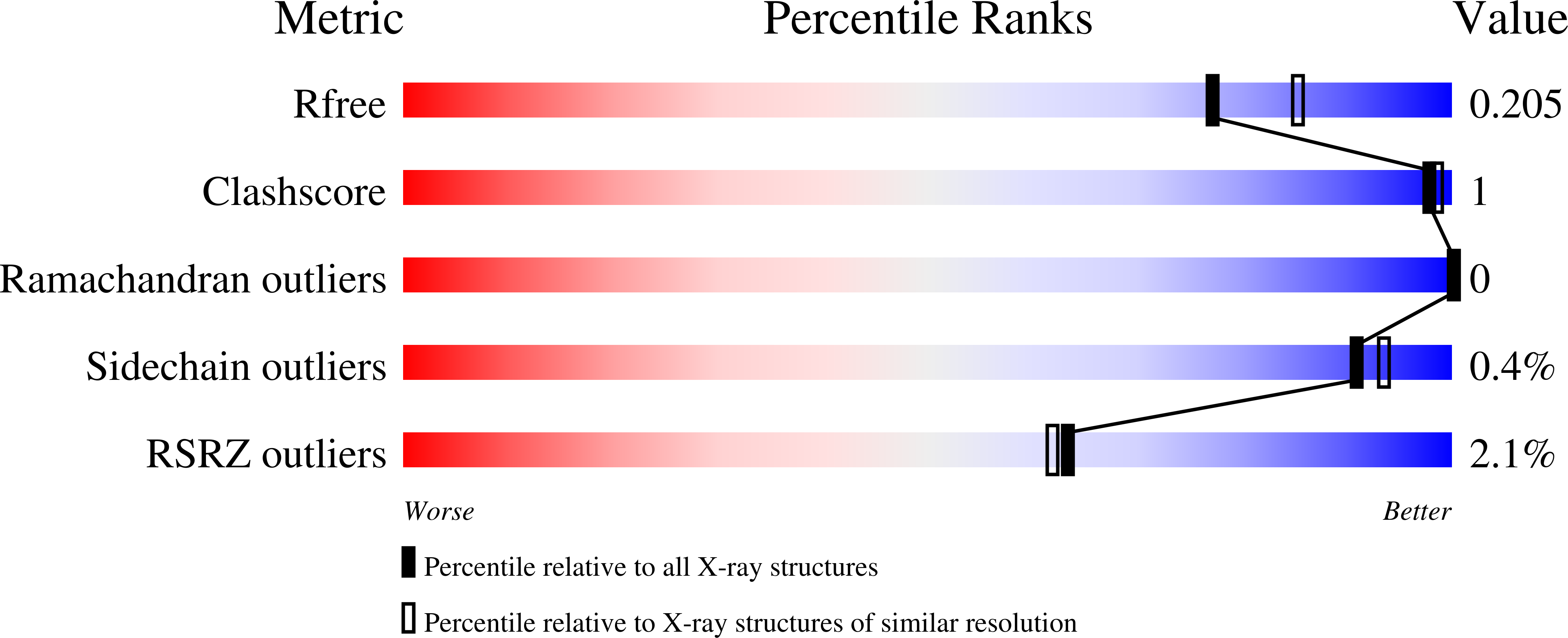
wwPDB Validation 3D Report Full Report
Entity ID: 1 | |||||
|---|---|---|---|---|---|
| Molecule | Chains | Sequence Length | Organism | Details | Image |
| Ribonucleoside-diphosphate reductase 2 subunit beta | 319 | Escherichia coli K-12 | Mutation(s): 0 Gene Names: b2676, JW2651, nrdF, ygaD EC: 1.17.4.1 |  | |
UniProt | |||||
Find proteins for P37146 (Escherichia coli (strain K12)) Explore P37146 Go to UniProtKB: P37146 | |||||
Entity Groups | |||||
| Sequence Clusters | 30% Identity50% Identity70% Identity90% Identity95% Identity100% Identity | ||||
| UniProt Group | P37146 | ||||
Sequence AnnotationsExpand | |||||
| |||||
| Length ( Å ) | Angle ( ˚ ) |
|---|---|
| a = 78.511 | α = 90 |
| b = 78.511 | β = 90 |
| c = 267.294 | γ = 120 |
| Software Name | Purpose |
|---|---|
| DENZO | data reduction |
| SCALEPACK | data scaling |
| REFMAC | refinement |
| PDB_EXTRACT | data extraction |
| HKL-2000 | data reduction |
| HKL-2000 | data scaling |
| PHASER | phasing |
RCSB PDB (citation) is hosted by
RCSB PDB is a member of the

