


Crystal structures of E.coli glutaredoxin 2 with glutathione and of a mutant C9S/C12S without glutathione
Ye, J., Venkadesh, S., Rosen, B.P.To be published.
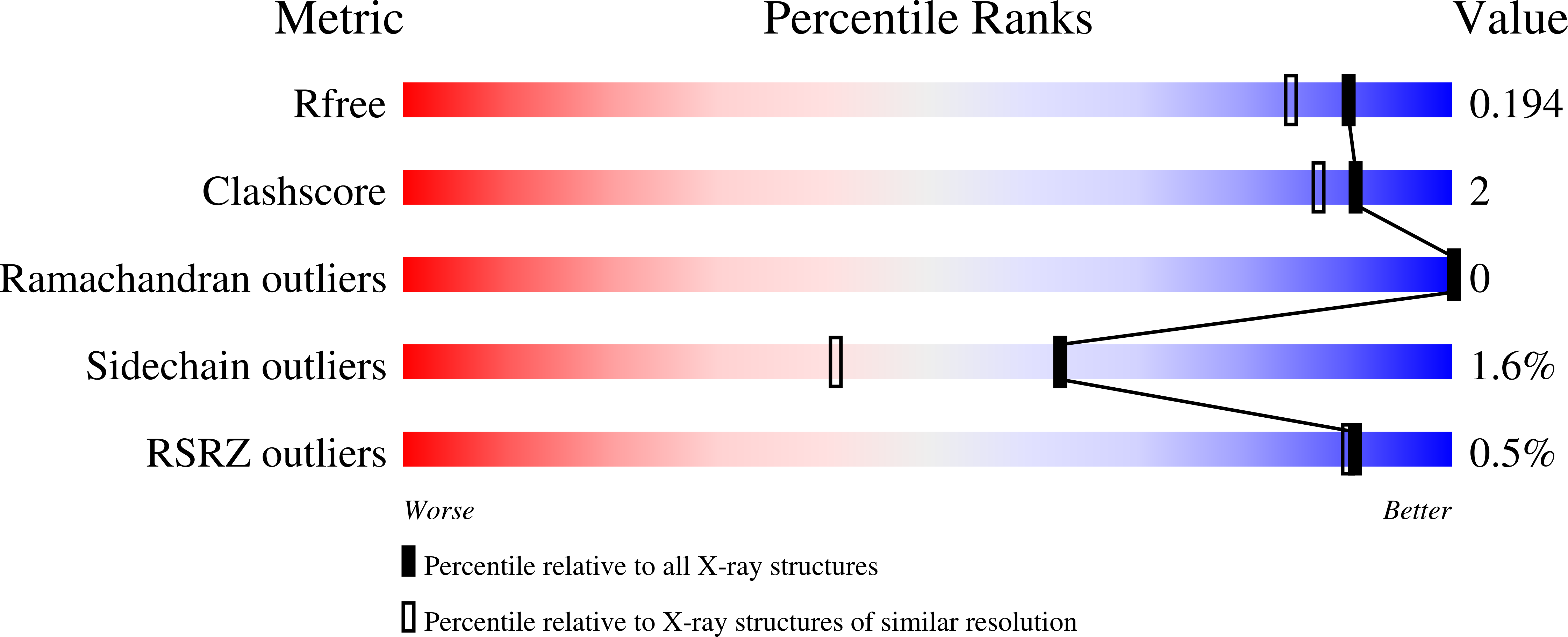
Experimental Data Snapshot
Entity ID: 1 | |||||
|---|---|---|---|---|---|
| Molecule | Chains | Sequence Length | Organism | Details | Image |
| Glutaredoxin-2 | 217 | Escherichia coli K-12 | Mutation(s): 0 Gene Names: grxB, b1064, JW1051 | 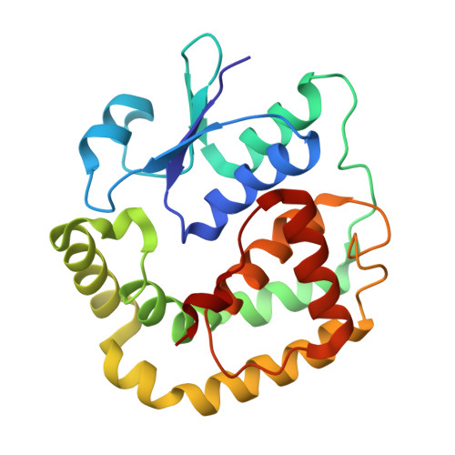 | |
UniProt | |||||
Find proteins for P0AC59 (Escherichia coli (strain K12)) Explore P0AC59 Go to UniProtKB: P0AC59 | |||||
Entity Groups | |||||
| Sequence Clusters | 30% Identity50% Identity70% Identity90% Identity95% Identity100% Identity | ||||
| UniProt Group | P0AC59 | ||||
Sequence AnnotationsExpand | |||||
| |||||
| Ligands 2 Unique | |||||
|---|---|---|---|---|---|
| ID | Chains | Name / Formula / InChI Key | 2D Diagram | 3D Interactions | |
| GSH Query on GSH | B [auth A] | GLUTATHIONE C10 H17 N3 O6 S RWSXRVCMGQZWBV-WDSKDSINSA-N |  | ||
| ACY Query on ACY | C [auth A] | ACETIC ACID C2 H4 O2 QTBSBXVTEAMEQO-UHFFFAOYSA-N |  | ||
| Length ( Å ) | Angle ( ˚ ) |
|---|---|
| a = 50.1 | α = 90 |
| b = 50.1 | β = 90 |
| c = 152.47 | γ = 120 |
| Software Name | Purpose |
|---|---|
| AMoRE | phasing |
| PHENIX | refinement |
| MOSFLM | data reduction |
| SCALA | data scaling |
RCSB PDB (citation) is hosted by
RCSB PDB is a member of the

