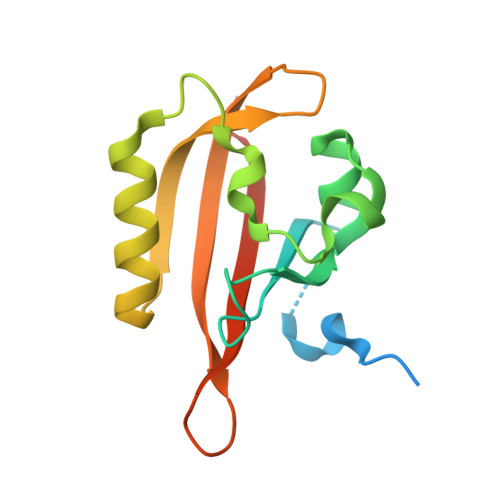
Structure and function of a short LOV protein from the marine phototrophic bacterium Dinoroseobacter shibae.
Endres, S., Granzin, J., Circolone, F., Stadler, A., Krauss, U., Drepper, T., Svensson, V., Knieps-Grunhagen, E., Wirtz, A., Cousin, A., Tielen, P., Willbold, D., Jaeger, K.E., Batra-Safferling, R.(2015) BMC Microbiol 15: 30-30
- PubMed: 25887755
- DOI: https://doi.org/10.1186/s12866-015-0365-0
- Primary Citation of Related Structures:
4KUK, 4KUO - PubMed Abstract:
Light, oxygen, voltage (LOV) domains are widely distributed in plants, algae, fungi, bacteria, and represent the photo-responsive domains of various blue-light photoreceptor proteins. Their photocycle involves the blue-light triggered adduct formation between the C(4a) atom of a non-covalently bound flavin chromophore and the sulfur atom of a conserved cysteine in the LOV sensor domain. LOV proteins show considerable variation in the structure of N- and C-terminal elements which flank the LOV core domain, as well as in the lifetime of the adduct state. Here, we report the photochemical, structural and functional characterization of DsLOV, a LOV protein from the photoheterotrophic marine α-proteobacterium Dinoroseobacter shibae which exhibits an average adduct state lifetime of 9.6 s at 20°C, and thus represents the fastest reverting bacterial LOV protein reported so far. Mutational analysis in D. shibae revealed a unique role of DsLOV in controlling the induction of photopigment synthesis in the absence of blue-light. The dark state crystal structure of DsLOV determined at 1.5 Å resolution reveals a conserved core domain with an extended N-terminal cap. The dimer interface in the crystal structure forms a unique network of hydrogen bonds involving residues of the N-terminus and the β-scaffold of the core domain. The structure of photoexcited DsLOV suggests increased flexibility in the N-cap region and a significant shift in the Cα backbone of β strands in the N- and C-terminal ends of the LOV core domain. The results presented here cover the characterization of the unusual short LOV protein DsLOV from Dinoroseobacter shibae including its regulatory function, extremely fast dark recovery and an N-terminus mediated dimer interface. Due to its unique photophysical, structural and regulatory properties, DsLOV might thus serve as an alternative model system for studying light perception by LOV proteins and physiological responses in bacteria.
Organizational Affiliation:
Institute of Molecular Enzyme Technology, Heinrich-Heine-Universität Düsseldorf, Forschungszentrum Jülich, D-52425, Jülich, Germany. s.endres@fz-juelich.de.