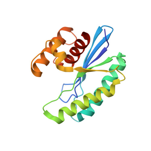
Mutants of phage bIL67 RuvC with enhanced Holliday junction binding selectivity and resolution symmetry.
Green, V., Curtis, F.A., Sedelnikova, S., Rafferty, J.B., Sharples, G.J.(2013) Mol Microbiol 89: 1240-1258
- PubMed: 23888987
- DOI: https://doi.org/10.1111/mmi.12343
- Primary Citation of Related Structures:
4KTW, 4KTZ - PubMed Abstract:
Viral and bacterial Holliday junction resolvases differ in specificity with the former typically being more promiscuous, acting on a variety of branched DNA substrates, while the latter exclusively targets Holliday junctions. We have determined the crystal structure of a RuvC resolvase from bacteriophage bIL67 to help identify features responsible for DNA branch discrimination. Comparisons between phage and bacterial RuvC structures revealed significant differences in the number and position of positively-charged residues in the outer sides of the junction binding cleft. Substitutions were generated in phage RuvC residues implicated in branch recognition and six were found to confer defects in Holliday junction and replication fork cleavage in vivo. Two mutants, R121A and R124A that flank the DNA binding site were purified and exhibited reduced in vitro binding to fork and linear duplex substrates relative to the wild-type, while retaining the ability to bind X junctions. Crucially, these two variants cleaved Holliday junctions with enhanced specificity and symmetry, a feature more akin to cellular RuvC resolvases. Thus, additional positive charges in the phage RuvC binding site apparently stabilize productive interactions with branched structures other than the canonical Holliday junction, a feature advantageous for viral DNA processing but deleterious for their cellular counterparts.
Organizational Affiliation:
Krebs Institute, Department of Molecular Biology and Biotechnology, University of Sheffield, Western Bank, Sheffield, S10 2TN, UK.