Structural and mutational analyses of Aes, an inhibitor of MalT in Escherichia coli.
Schiefner, A., Gerber, K., Brosig, A., Boos, W.(2014) Proteins 82: 268-277
- PubMed: 23934774
- DOI: https://doi.org/10.1002/prot.24383
- Primary Citation of Related Structures:
4KRX, 4KRY - PubMed Abstract:
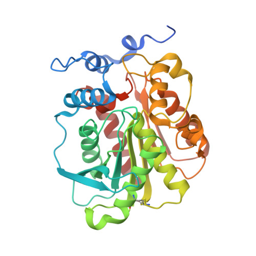
The acyl esterase Aes effectively inhibits the transcriptional activity of MalT-the central activator of maltose and maltodextrin utilizing genes in Escherichia coli. To provide better insight into the nature of the interaction between Aes and MalT, we determined two different crystal structures of Aes-in its native form and covalently modified by a phenylmethylsulfonyl moiety at its active site serine. Both structures show distinct space groups and were refined to a resolution of 1.8 Å and 2.3 Å, respectively. The overall structure of Aes resembles a canonical α/β-hydrolase fold, which is extended by a funnel-like cap structure that forms the substrate-binding site. The catalytic triad of Aes, comprising residues Ser165, His292, and Asp262, is located at the bottom of this funnel. Analysis of the crystal-packing contacts of the two different space groups as well as analytical size-exclusion chromatography revealed a homodimeric arrangement of Aes. The Aes dimer adopts an antiparallel contact involving both the hydrolase core and the cap, with its twofold axis perpendicular to the largest dimension of Aes. To identify the surface area of Aes that is responsible for the interaction with MalT, we performed a structure-based alanine-scanning mutagenesis to pinpoint Aes residues that are significantly impaired in MalT inhibition, but still exhibit wild-type expression and enzymatic activity. These residues map to a shallow slightly concave surface patch of Aes at the opposite site of the dimerization interface and indicate the surface area that interacts with MalT.
Organizational Affiliation:
Lehrstuhl für Biologische Chemie, Technische Universität München, 85350, Freising-Weihenstephan, Germany.