4KKI

Crystal Structure of Haptocorrin in Complex with CNCbl


Experimental Data Snapshot

  • Method: X-RAY DIFFRACTION
  • Resolution: 2.35 Å
  • R-Value Free: 0.226 
  • R-Value Work: 0.196 
  • R-Value Observed: 0.197 

wwPDB Validation   3D Report Full Report


Ligand Structure Quality Assessment 


This is version 2.1 of the entry. See complete history


Literature

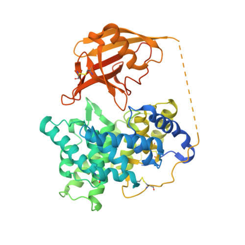
Structural basis for universal corrinoid recognition by the cobalamin transport protein haptocorrin.

Furger, E.Frei, D.C.Schibli, R.Fischer, E.Prota, A.E.

(2013) J Biol Chem 288: 25466-25476

  • DOI: https://doi.org/10.1074/jbc.M113.483271
  • Primary Citation of Related Structures:  
    4KKI, 4KKJ

  • PubMed Abstract: 

    Cobalamin (Cbl; vitamin B12) is an essential micronutrient synthesized only by bacteria. Mammals have developed a sophisticated uptake system to capture the vitamin from the diet. Cbl transport is mediated by three transport proteins: transcobalamin, intrinsic factor, and haptocorrin (HC). All three proteins have a similar overall structure but a different selectivity for corrinoids. Here, we present the crystal structures of human HC in complex with cyanocobalamin and cobinamide at 2.35 and 3.0 Å resolution, respectively. The structures reveal that many of the interactions with the corrin ring are conserved among the human Cbl transporters. However, the non-conserved residues Asn-120, Arg-357, and Asn-373 form distinct interactions allowing for stabilization of corrinoids other than Cbl. A central binding motif forms interactions with the e- and f-side chains of the corrin ring and is conserved in corrinoid-binding proteins of other species. In addition, the α- and β-domains of HC form several unique interdomain contacts and have a higher shape complementarity than those of intrinsic factor and transcobalamin. The stabilization of ligands by all of these interactions is reflected in higher melting temperatures of the protein-ligand complexes. Our structural analysis offers fundamental insights into the unique binding behavior of HC and completes the picture of Cbl interaction with its three transport proteins.


  • Organizational Affiliation

    From the Center for Radiopharmaceutical Sciences and.


Macromolecules
Find similar proteins by:  (by identity cutoff)  |  3D Structure
Entity ID: 1
MoleculeChains Sequence LengthOrganismDetailsImage
Transcobalamin-1467Homo sapiensMutation(s): 0 
Gene Names: TC1TCN1
UniProt & NIH Common Fund Data Resources
Find proteins for P20061 (Homo sapiens)
Explore P20061 
Go to UniProtKB:  P20061
PHAROS:  P20061
GTEx:  ENSG00000134827 
Entity Groups  
Sequence Clusters30% Identity50% Identity70% Identity90% Identity95% Identity100% Identity
UniProt GroupP20061
Sequence Annotations
Expand
  • Reference Sequence
Small Molecules
Ligands 4 Unique
IDChains Name / Formula / InChI Key2D Diagram3D Interactions
CNC
Query on CNC

Download Ideal Coordinates CCD File 
I [auth A]CYANOCOBALAMIN
C63 H89 Co N14 O14 P
SYZBZQWSWIJYAR-UVKKECPRSA-M
NAG
Query on NAG

Download Ideal Coordinates CCD File 
B [auth A]
C [auth A]
D [auth A]
E [auth A]
F [auth A]
B [auth A],
C [auth A],
D [auth A],
E [auth A],
F [auth A],
G [auth A],
H [auth A]
2-acetamido-2-deoxy-beta-D-glucopyranose
C8 H15 N O6
OVRNDRQMDRJTHS-FMDGEEDCSA-N
PEG
Query on PEG

Download Ideal Coordinates CCD File 
K [auth A]
L [auth A]
M [auth A]
N [auth A]
O [auth A]
K [auth A],
L [auth A],
M [auth A],
N [auth A],
O [auth A],
P [auth A],
Q [auth A],
R [auth A],
S [auth A],
T [auth A],
U [auth A]
DI(HYDROXYETHYL)ETHER
C4 H10 O3
MTHSVFCYNBDYFN-UHFFFAOYSA-N
CA
Query on CA

Download Ideal Coordinates CCD File 
J [auth A]CALCIUM ION
Ca
BHPQYMZQTOCNFJ-UHFFFAOYSA-N
Experimental Data & Validation

Experimental Data

  • Method: X-RAY DIFFRACTION
  • Resolution: 2.35 Å
  • R-Value Free: 0.226 
  • R-Value Work: 0.196 
  • R-Value Observed: 0.197 
  • Space Group: P 64
Unit Cell:
Length ( Å )Angle ( ˚ )
a = 149.89α = 90
b = 149.89β = 90
c = 57.49γ = 120
Software Package:
Software NamePurpose
RemDAqdata collection
PHASERphasing
PHENIXrefinement
XDSdata reduction
XDSdata scaling

Structure Validation

View Full Validation Report


Ligand Structure Quality Assessment 


Entry History 

Deposition Data

Revision History  (Full details and data files)

  • Version 1.0: 2013-07-17
    Type: Initial release
  • Version 1.1: 2013-10-09
    Changes: Database references
  • Version 1.2: 2017-11-15
    Changes: Refinement description
  • Version 1.3: 2020-07-29
    Type: Remediation
    Reason: Carbohydrate remediation
    Changes: Data collection, Database references, Derived calculations, Structure summary
  • Version 2.0: 2021-08-18
    Changes: Data collection, Database references, Derived calculations, Non-polymer description, Structure summary
  • Version 2.1: 2023-09-20
    Changes: Data collection, Refinement description