4K6O

X-ray structure uridine phosphorylase from Vibrio cholerae in complex with 6-methyluracil at 1.17 A resolution


Experimental Data Snapshot

  • Method: X-RAY DIFFRACTION
  • Resolution: 1.17 Å
  • R-Value Free: 0.128 
  • R-Value Work: 0.108 
  • R-Value Observed: 0.110 

wwPDB Validation   3D Report Full Report


This is version 1.3 of the entry. See complete history


Literature

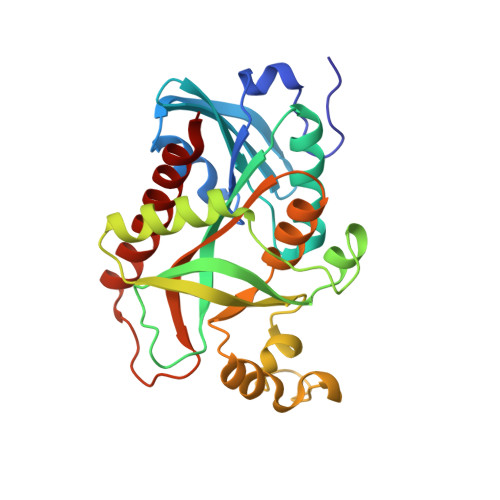
Crystallization and preliminary X-ray study of Vibrio cholerae uridine phosphorylase in complex with 6-methyluracil.

Prokofev, I.I.Lashkov, A.A.Gabdulkhakov, A.G.Dontsova, M.V.Seregina, T.A.Mironov, A.S.Betzel, C.Mikhailov, A.M.

(2014) Acta Crystallogr F Struct Biol Commun 70: 60-63

  • DOI: https://doi.org/10.1107/S2053230X13031877
  • Primary Citation of Related Structures:  
    4K6O

  • PubMed Abstract: 

    Uridine phosphorylase catalyzes the phosphorolysis of ribonucleosides, with the nitrogenous base and ribose 1-phosphate as products. Additionally, it catalyzes the reverse reaction of the synthesis of ribonucleosides from ribose 1-phosphate and a nitrogenous base. However, the enzyme does not catalyze the synthesis of nucleosides when the substrate is a nitrogenous base substituted at the 6-position, such as 6-methyluracil (6-MU). In order to explain this fact, it is essential to investigate the three-dimensional structure of the complex of 6-MU with uridine phosphorylase. 6-MU is a pharmaceutical agent that improves tissue nutrition and enhances cell regeneration by normalization of nucleotide exchange in humans. 6-MU is used for the treatment of diseases of the gastrointestinal tract, including infectious diseases. Here, procedures to obtain the uridine phosphorylase from the pathogenic bacterium Vibrio cholerae (VchUPh), purification of this enzyme, crystallization of the complex of VchUPh with 6-MU, and X-ray data collection and preliminary X-ray analysis of the VchUPh-6-MU complex at atomic resolution are reported.


  • Organizational Affiliation

    A. V. Shubnikov Institute of Crystallography, Russian Academy of Sciences, Leninsky Prospect 59, Moscow 117333, Russian Federation.


Macromolecules
Find similar proteins by:  (by identity cutoff)  |  3D Structure
Entity ID: 1
MoleculeChains Sequence LengthOrganismDetailsImage
Uridine phosphorylase
A, B, C, D, E
A, B, C, D, E, F
253Vibrio choleraeMutation(s): 0 
Gene Names: udp
EC: 2.4.2.3
UniProt
Find proteins for Q9K4U1 (Vibrio cholerae)
Explore Q9K4U1 
Go to UniProtKB:  Q9K4U1
Entity Groups  
Sequence Clusters30% Identity50% Identity70% Identity90% Identity95% Identity100% Identity
UniProt GroupQ9K4U1
Sequence Annotations
Expand
  • Reference Sequence
Small Molecules
Ligands 8 Unique
IDChains Name / Formula / InChI Key2D Diagram3D Interactions
6MU
Query on 6MU

Download Ideal Coordinates CCD File 
CA [auth C]
CB [auth F]
I [auth A]
MA [auth D]
R [auth B]
CA [auth C],
CB [auth F],
I [auth A],
MA [auth D],
R [auth B],
TA [auth E]
6-methylpyrimidine-2,4-diol
C5 H6 N2 O2
SHVCSCWHWMSGTE-UHFFFAOYSA-N
TRS
Query on TRS

Download Ideal Coordinates CCD File 
HA [auth C],
W [auth B],
WA [auth E],
XA [auth E]
2-AMINO-2-HYDROXYMETHYL-PROPANE-1,3-DIOL
C4 H12 N O3
LENZDBCJOHFCAS-UHFFFAOYSA-O
GOL
Query on GOL

Download Ideal Coordinates CCD File 
KA [auth C]GLYCEROL
C3 H8 O3
PEDCQBHIVMGVHV-UHFFFAOYSA-N
EDO
Query on EDO

Download Ideal Coordinates CCD File 
GB [auth F],
M [auth A],
QA [auth D],
X [auth B]
1,2-ETHANEDIOL
C2 H6 O2
LYCAIKOWRPUZTN-UHFFFAOYSA-N
EOH
Query on EOH

Download Ideal Coordinates CCD File 
HB [auth F]
IA [auth C]
JA [auth C]
N [auth A]
RA [auth D]
HB [auth F],
IA [auth C],
JA [auth C],
N [auth A],
RA [auth D],
Y [auth B],
YA [auth E]
ETHANOL
C2 H6 O
LFQSCWFLJHTTHZ-UHFFFAOYSA-N
CL
Query on CL

Download Ideal Coordinates CCD File 
AA [auth C]
AB [auth F]
BA [auth C]
BB [auth F]
G [auth A]
AA [auth C],
AB [auth F],
BA [auth C],
BB [auth F],
G [auth A],
H [auth A],
LA [auth D],
O [auth A],
P [auth B],
Q [auth B],
SA [auth E],
Z [auth C]
CHLORIDE ION
Cl
VEXZGXHMUGYJMC-UHFFFAOYSA-M
MG
Query on MG

Download Ideal Coordinates CCD File 
DA [auth C]
DB [auth F]
EA [auth C]
EB [auth F]
FA [auth C]
DA [auth C],
DB [auth F],
EA [auth C],
EB [auth F],
FA [auth C],
FB [auth F],
J [auth A],
K [auth A],
NA [auth D],
OA [auth D],
PA [auth D],
S [auth B],
T [auth B],
U [auth B],
UA [auth E],
ZA [auth E]
MAGNESIUM ION
Mg
JLVVSXFLKOJNIY-UHFFFAOYSA-N
NA
Query on NA

Download Ideal Coordinates CCD File 
GA [auth C],
L [auth A],
V [auth B],
VA [auth E]
SODIUM ION
Na
FKNQFGJONOIPTF-UHFFFAOYSA-N
Experimental Data & Validation

Experimental Data

  • Method: X-RAY DIFFRACTION
  • Resolution: 1.17 Å
  • R-Value Free: 0.128 
  • R-Value Work: 0.108 
  • R-Value Observed: 0.110 
  • Space Group: P 1 21 1
Unit Cell:
Length ( Å )Angle ( ˚ )
a = 93.012α = 90
b = 97.1β = 119.99
c = 93.017γ = 90
Software Package:
Software NamePurpose
XSCALEdata scaling
MOLREPphasing
PHENIXrefinement
PDB_EXTRACTdata extraction

Structure Validation

View Full Validation Report


Entry History 

Deposition Data

Revision History  (Full details and data files)

  • Version 1.0: 2014-05-07
    Type: Initial release
  • Version 1.1: 2014-05-14
    Changes: Data collection, Database references
  • Version 1.2: 2019-12-18
    Changes: Data collection, Database references
  • Version 1.3: 2023-11-08
    Changes: Data collection, Database references, Derived calculations, Refinement description