


Crystal structure of an enoyl-CoA hydratase/isomerase from Marinobacter aquaeolei
Eswaramoorthy, S., Almo, S.C., Swaminathan, S.To be published.
Experimental Data Snapshot
wwPDB Validation 3D Report Full Report

Entity ID: 1 | |||||
|---|---|---|---|---|---|
| Molecule | Chains | Sequence Length | Organism | Details | Image |
| Enoyl-CoA hydratase/isomerase | 296 | Marinobacter nauticus VT8 | Mutation(s): 0 Gene Names: Maqu_2806 | 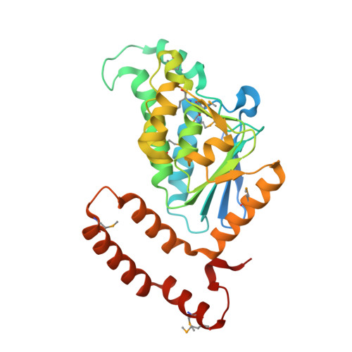 | |
UniProt | |||||
Find proteins for A1U4G2 (Marinobacter nauticus (strain ATCC 700491 / DSM 11845 / VT8)) Explore A1U4G2 Go to UniProtKB: A1U4G2 | |||||
Entity Groups | |||||
| Sequence Clusters | 30% Identity50% Identity70% Identity90% Identity95% Identity100% Identity | ||||
| UniProt Group | A1U4G2 | ||||
Sequence AnnotationsExpand | |||||
| |||||
| Modified Residues 1 Unique | |||||
|---|---|---|---|---|---|
| ID | Chains | Type | Formula | 2D Diagram | Parent |
| MSE Query on MSE | A, B, C | L-PEPTIDE LINKING | C5 H11 N O2 Se |  | MET |
| Length ( Å ) | Angle ( ˚ ) |
|---|---|
| a = 232.355 | α = 90 |
| b = 135.143 | β = 99.09 |
| c = 43.042 | γ = 90 |
| Software Name | Purpose |
|---|---|
| CBASS | data collection |
| SHELXS | phasing |
| REFMAC | refinement |
| HKL-2000 | data reduction |
| HKL-2000 | data scaling |
RCSB PDB (citation) is hosted by
RCSB PDB is a member of the

