4JYO

Structural basis for angiopoietin-1 mediated signaling initiation


Experimental Data Snapshot

  • Method: X-RAY DIFFRACTION
  • Resolution: 2.50 Å
  • R-Value Free: 0.242 
  • R-Value Work: 0.186 
  • R-Value Observed: 0.189 

wwPDB Validation   3D Report Full Report


This is version 1.2 of the entry. See complete history


Literature

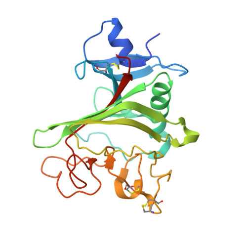
Structural basis for angiopoietin-1-mediated signaling initiation.

Yu, X.Seegar, T.C.Dalton, A.C.Tzvetkova-Robev, D.Goldgur, Y.Rajashankar, K.R.Nikolov, D.B.Barton, W.A.

(2013) Proc Natl Acad Sci U S A 110: 7205-7210

  • DOI: https://doi.org/10.1073/pnas.1216890110
  • Primary Citation of Related Structures:  
    4JYO, 4JZC, 4K0V

  • PubMed Abstract: 

    Angiogenesis is a complex cellular process involving multiple regulatory growth factors and growth factor receptors. Among them, the ligands for the endothelial-specific tunica intima endothelial receptor tyrosine kinase 2 (Tie2) receptor kinase, angiopoietin-1 (Ang1) and Ang2, play essential roles in balancing vessel stability and regression during both developmental and tumor-induced angiogenesis. Despite possessing a high degree of sequence identity, Ang1 and Ang2 have distinct functional roles and cell-signaling characteristics. Here, we present the crystal structures of Ang1 both unbound and in complex with the Tie2 ectodomain. Comparison of the Ang1-containing structures with their Ang2-containing counterparts provide insight into the mechanism of receptor activation and reveal molecular surfaces important for interactions with Tie2 coreceptors and associated signaling proteins. Using structure-based mutagenesis, we identify a loop within the angiopoietin P domain, adjacent to the receptor-binding interface, which confers the specific agonist/antagonist properties of the molecule. We demonstrate using cell-based assays that an Ang2 chimera containing the Ang1 loop sequence behaves functionally similarly to Ang1 as a constitutive Tie2 agonist, able to efficiently dissociate the inhibitory Tie1/Tie2 complex and elicit Tie2 clustering and downstream signaling.


  • Organizational Affiliation

    Structural Biology Program, Memorial Sloan-Kettering Cancer Center, New York, NY 10021, USA.


Macromolecules
Find similar proteins by:  (by identity cutoff)  |  3D Structure
Entity ID: 1
MoleculeChains Sequence LengthOrganismDetailsImage
Angiopoietin-1A [auth X]230Homo sapiensMutation(s): 0 
Gene Names: ANGPT1KIAA0003
UniProt & NIH Common Fund Data Resources
Find proteins for Q15389 (Homo sapiens)
Explore Q15389 
Go to UniProtKB:  Q15389
PHAROS:  Q15389
GTEx:  ENSG00000154188 
Entity Groups  
Sequence Clusters30% Identity50% Identity70% Identity90% Identity95% Identity100% Identity
UniProt GroupQ15389
Sequence Annotations
Expand
  • Reference Sequence
Small Molecules
Ligands 1 Unique
IDChains Name / Formula / InChI Key2D Diagram3D Interactions
CA
Query on CA

Download Ideal Coordinates CCD File 
B [auth X]CALCIUM ION
Ca
BHPQYMZQTOCNFJ-UHFFFAOYSA-N
Experimental Data & Validation

Experimental Data

  • Method: X-RAY DIFFRACTION
  • Resolution: 2.50 Å
  • R-Value Free: 0.242 
  • R-Value Work: 0.186 
  • R-Value Observed: 0.189 
  • Space Group: P 64 2 2
Unit Cell:
Length ( Å )Angle ( ˚ )
a = 80.968α = 90
b = 80.968β = 90
c = 187.216γ = 120
Software Package:
Software NamePurpose
ADSCdata collection
AMoREphasing
REFMACrefinement
HKL-2000data reduction
HKL-2000data scaling

Structure Validation

View Full Validation Report


Entry History 

Deposition Data

Revision History  (Full details and data files)

  • Version 1.0: 2013-05-08
    Type: Initial release
  • Version 1.1: 2013-05-15
    Changes: Database references
  • Version 1.2: 2023-09-20
    Changes: Data collection, Database references, Derived calculations, Refinement description