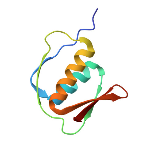
A dodecameric ring-like structure of the N0 domain of the type II secretin from enterotoxigenic Escherichia coli.
Korotkov, K.V., Delarosa, J.R., Hol, W.G.(2013) J Struct Biol 18: 354-362
- PubMed: 23820381
- DOI: https://doi.org/10.1016/j.jsb.2013.06.013
- Primary Citation of Related Structures:
4JTM - PubMed Abstract:
In many bacteria, secretins from the type II secretion system (T2SS) function as outer membrane gated channels that enable passage of folded proteins from the periplasm into the extracellular milieu. Cryo-electron microscopy of the T2SS secretin GspD revealed previously the dodecameric cylindrical architecture of secretins, and crystal structures of periplasmic secretin domains showed a modular domain organization. However, no high-resolution experimental data has as yet been provided about how the entire T2SS secretin or its domains are organized in a cylindrical fashion. Here we present a crystal structure of the N0 domain of the T2SS secretin GspD from enterotoxigenic Escherichia coli containing a helix with 12 subunits per turn. The helix has an outer diameter of ∼125Å and a pitch of only 24Å which suggests a model of a cylindrical dodecameric N0 ring whose dimensions correspond with the cryo-electron microscopy map of Vibrio cholerae GspD. The N0 domain is known to interact with the HR domain of the inner membrane T2SS protein GspC. When the new N0 ring model is combined with the known N0·HR crystal structure, a dodecameric double-ring of twelve N0-HR heterodimers is obtained. In contrast, the previously observed compact N0-N1 GspD module is not compatible with the N0 ring. Interestingly, a N0-N1 T3SS homolog is compatible with forming a N0-N1 dodecameric ring, due to a different N0-vs-N1 orientation. This suggests that the dodecameric N0 ring is an important feature of T2SS secretins with periplasmic domains undergoing considerable motions during exoprotein translocation.
Organizational Affiliation:
Department of Biochemistry and Biomolecular Structure Center, University of Washington, Seattle, WA 98195, United States.