


X-ray structure of uridine phosphorylase from Yersinia pseudotuberculosis in unliganded state at 2.27 A resolution
Balaev, V.V., Lashkov, A.A., Prokofev, I.I., Gabdoulkhakov, A.G., Betzel, C., Mikhailov, A.M.To be published.
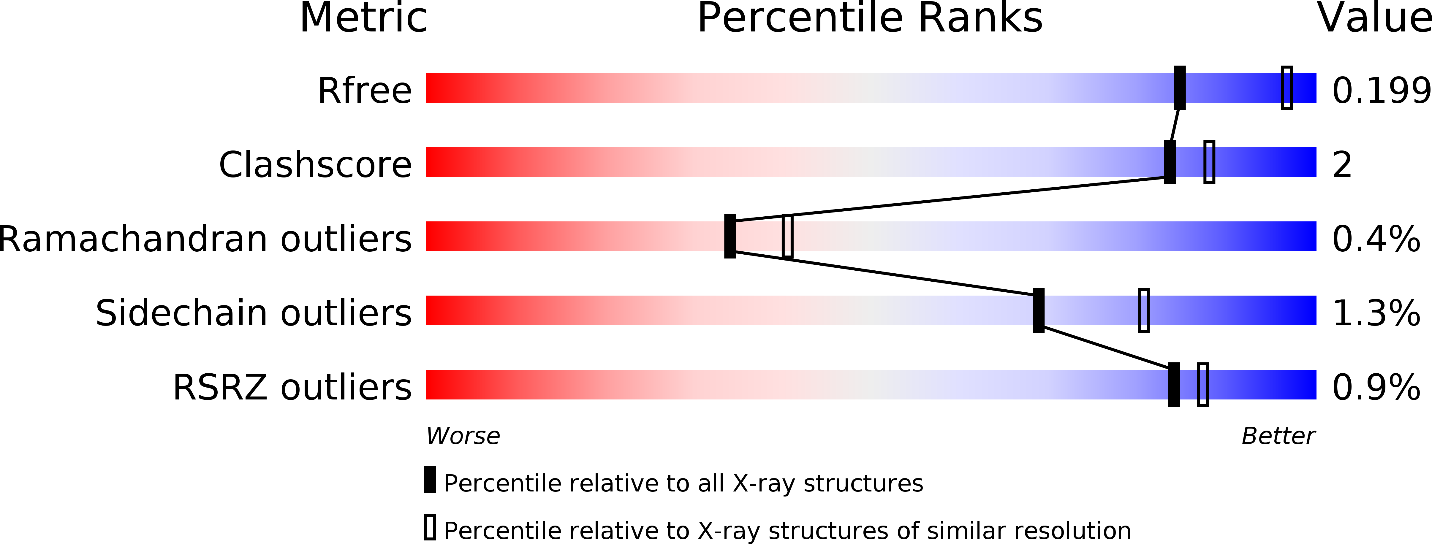
Experimental Data Snapshot
wwPDB Validation 3D Report Full Report
Entity ID: 1 | |||||
|---|---|---|---|---|---|
| Molecule | Chains | Sequence Length | Organism | Details | Image |
| Uridine phosphorylase | 253 | Yersinia pestis | Mutation(s): 0 Gene Names: udp, y0444, YP_3263 EC: 2.4.2.3 | 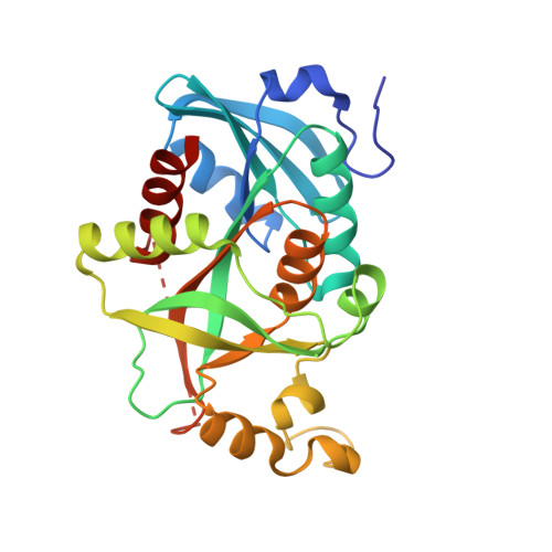 | |
UniProt | |||||
Find proteins for Q8D1I4 (Yersinia pestis) Explore Q8D1I4 Go to UniProtKB: Q8D1I4 | |||||
Entity Groups | |||||
| Sequence Clusters | 30% Identity50% Identity70% Identity90% Identity95% Identity100% Identity | ||||
| UniProt Group | Q8D1I4 | ||||
Sequence AnnotationsExpand | |||||
| |||||
| Ligands 3 Unique | |||||
|---|---|---|---|---|---|
| ID | Chains | Name / Formula / InChI Key | 2D Diagram | 3D Interactions | |
| SO4 Query on SO4 | H [auth C] | SULFATE ION O4 S QAOWNCQODCNURD-UHFFFAOYSA-L |  | ||
| GOL Query on GOL | I [auth E], J [auth E] | GLYCEROL C3 H8 O3 PEDCQBHIVMGVHV-UHFFFAOYSA-N |  | ||
| EDO Query on EDO | G [auth A], K [auth F], L [auth F] | 1,2-ETHANEDIOL C2 H6 O2 LYCAIKOWRPUZTN-UHFFFAOYSA-N |  | ||
| Length ( Å ) | Angle ( ˚ ) |
|---|---|
| a = 93.75 | α = 90 |
| b = 93.75 | β = 90 |
| c = 146.58 | γ = 120 |
| Software Name | Purpose |
|---|---|
| XSCALE | data scaling |
| REFMAC | refinement |
| PDB_EXTRACT | data extraction |
| MxCuBE | data collection |
| XDS | data reduction |
| MOLREP | phasing |
RCSB PDB (citation) is hosted by
RCSB PDB is a member of the

