4JFU

Crystal structure of a bacterial fucosidase with iminosugar inhibitor


Experimental Data Snapshot

  • Method: X-RAY DIFFRACTION
  • Resolution: 1.66 Å
  • R-Value Free: 0.177 
  • R-Value Work: 0.152 
  • R-Value Observed: 0.153 

wwPDB Validation   3D Report Full Report


Ligand Structure Quality Assessment 


This is version 1.1 of the entry. See complete history


Literature

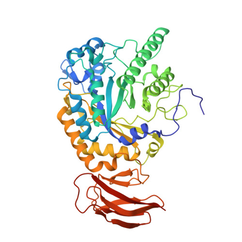
alpha-L-fucosidase inhibition by pyrrolidine-ferrocene hybrids: rationalization of ligand-binding properties by structural studies.

Hottin, A.Wright, D.W.Steenackers, A.Delannoy, P.Dubar, F.Biot, C.Davies, G.J.Behr, J.B.

(2013) Chemistry 19: 9526-9533

  • DOI: https://doi.org/10.1002/chem.201301001
  • Primary Citation of Related Structures:  
    4JFS, 4JFT, 4JFU, 4JFV, 4JFW

  • PubMed Abstract: 

    Enhanced metabolism of fucose through fucosidase overexpression is a signature of some cancer types, thus suggesting that fucosidase-targetted ligands could play the role of drug-delivery vectors. Herein, we describe the synthesis of a new series of pyrrolidine-ferrocene conjugates, consisting of a L-fuco-configured dihydroxypyrrolidine as the fucosidase ligand armed with a cytotoxic ferrocenylamine moeity. Three-dimensional structures of several of these fucosidase inhibitors reveal transition-state-mimicking (3)E conformations. Elaboration with the ferrocenyl moiety results in sub-micromolar inhibitors of both bovine and bacterial fucosidases, with the 3D structure of the latter revealing electron density indicative of highly mobile alkylferrocene compounds. The best compounds show a strong antiproliferative effect, with up to 100% inhibition of the proliferation of MDA-MB-231 cancer cells at 50 μM.


  • Organizational Affiliation

    Université de Reims Champagne-Ardenne, Institut de Chimie Moléculaire de Reims, CNRS UMR 7312, UFR des Sciences Exactes et Naturelles, 51687 Reims Cedex 2, France.


Macromolecules
Find similar proteins by:  (by identity cutoff)  |  3D Structure
Entity ID: 1
MoleculeChains Sequence LengthOrganismDetailsImage
alpha-L-fucosidase
A, B
450Bacteroides thetaiotaomicron VPI-5482Mutation(s): 0 
UniProt
Find proteins for Q8A3I4 (Bacteroides thetaiotaomicron (strain ATCC 29148 / DSM 2079 / JCM 5827 / CCUG 10774 / NCTC 10582 / VPI-5482 / E50))
Explore Q8A3I4 
Go to UniProtKB:  Q8A3I4
Entity Groups  
Sequence Clusters30% Identity50% Identity70% Identity90% Identity95% Identity100% Identity
UniProt GroupQ8A3I4
Sequence Annotations
Expand
  • Reference Sequence
Small Molecules
Ligands 4 Unique
IDChains Name / Formula / InChI Key2D Diagram3D Interactions
K80
Query on K80

Download Ideal Coordinates CCD File 
C [auth A],
L [auth B]
(2S,3R,4S,5S)-2-methyl-5-(4-methylphenyl)pyrrolidine-3,4-diol
C12 H17 N O2
VSLJKQDKGOXAQN-IXLVHKGHSA-N
SO4
Query on SO4

Download Ideal Coordinates CCD File 
D [auth A],
E [auth A],
M [auth B]
SULFATE ION
O4 S
QAOWNCQODCNURD-UHFFFAOYSA-L
GOL
Query on GOL

Download Ideal Coordinates CCD File 
H [auth A],
I [auth A],
J [auth A],
K [auth A],
Q [auth B]
GLYCEROL
C3 H8 O3
PEDCQBHIVMGVHV-UHFFFAOYSA-N
IMD
Query on IMD

Download Ideal Coordinates CCD File 
F [auth A],
G [auth A],
N [auth B],
O [auth B],
P [auth B]
IMIDAZOLE
C3 H5 N2
RAXXELZNTBOGNW-UHFFFAOYSA-O
Experimental Data & Validation

Experimental Data

  • Method: X-RAY DIFFRACTION
  • Resolution: 1.66 Å
  • R-Value Free: 0.177 
  • R-Value Work: 0.152 
  • R-Value Observed: 0.153 
  • Space Group: P 1 21 1
Unit Cell:
Length ( Å )Angle ( ˚ )
a = 68.03α = 90
b = 95.4β = 90.83
c = 96.93γ = 90
Software Package:
Software NamePurpose
SCALAdata scaling
REFMACrefinement
PDB_EXTRACTdata extraction
MOSFLMdata reduction
REFMACphasing

Structure Validation

View Full Validation Report


Ligand Structure Quality Assessment 


Entry History 

Deposition Data

Revision History  (Full details and data files)

  • Version 1.0: 2013-09-18
    Type: Initial release
  • Version 1.1: 2023-09-20
    Changes: Data collection, Database references, Derived calculations, Refinement description