4JEC

Joint neutron and X-ray structure of per-deuterated HIV-1 protease in complex with clinical inhibitor amprenavir


Experimental Data Snapshot

  • Method: NEUTRON DIFFRACTION
  • Resolution: 2.00 Å
  • R-Value Free: 0.261 
  • R-Value Work: 0.244 
  • R-Value Observed: 0.244 

  • Method: X-RAY DIFFRACTION
  • Resolution: 2.01 Å
  • R-Value Free: 0.203 
  • R-Value Work: 0.194 

wwPDB Validation   3D Report Full Report


This is version 1.2 of the entry. See complete history


Literature

Joint X-ray/Neutron Crystallographic Study of HIV-1 Protease with Clinical Inhibitor Amprenavir: Insights for Drug Design.

Weber, I.T.Waltman, M.J.Mustyakimov, M.Blakeley, M.P.Keen, D.A.Ghosh, A.K.Langan, P.Kovalevsky, A.Y.

(2013) J Med Chem 56: 5631-5635

  • DOI: https://doi.org/10.1021/jm400684f
  • Primary Citation of Related Structures:  
    4JEC

  • PubMed Abstract: 

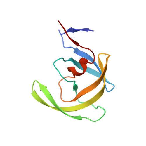
    HIV-1 protease is an important target for the development of antiviral inhibitors to treat AIDS. A room-temperature joint X-ray/neutron structure of the protease in complex with clinical drug amprenavir has been determined at 2.0 Å resolution. The structure provides direct determination of hydrogen atom positions in the enzyme active site. Analysis of the enzyme-drug interactions suggests that some hydrogen bonds may be weaker than deduced from the non-hydrogen interatomic distances. This information may be valuable for the design of improved protease inhibitors.


  • Organizational Affiliation

    Departments of Chemistry and Biology, Georgia State University , Atlanta, Georgia, United States.


Macromolecules
Find similar proteins by:  (by identity cutoff)  |  3D Structure
Entity ID: 1
MoleculeChains Sequence LengthOrganismDetailsImage
HIV-1 protease
A, B
99Human immunodeficiency virus 1Mutation(s): 5 
Gene Names: gag-pol
EC: 3.4.23.16
UniProt
Find proteins for P03367 (Human immunodeficiency virus type 1 group M subtype B (isolate BRU/LAI))
Explore P03367 
Go to UniProtKB:  P03367
Entity Groups  
Sequence Clusters30% Identity50% Identity70% Identity90% Identity95% Identity100% Identity
UniProt GroupP03367
Sequence Annotations
Expand
  • Reference Sequence
Small Molecules
Ligands 2 Unique
IDChains Name / Formula / InChI Key2D Diagram3D Interactions
478
Query on 478

Download Ideal Coordinates CCD File 
D [auth B]{3-[(4-AMINO-BENZENESULFONYL)-ISOBUTYL-AMINO]-1-BENZYL-2-HYDROXY-PROPYL}-CARBAMIC ACID TETRAHYDRO-FURAN-3-YL ESTER
C25 H35 N3 O6 S
YMARZQAQMVYCKC-OEMFJLHTSA-N
CL
Query on CL

Download Ideal Coordinates CCD File 
C [auth A]CHLORIDE ION
Cl
VEXZGXHMUGYJMC-UHFFFAOYSA-M
Binding Affinity Annotations 
IDSourceBinding Affinity
478 BindingDB:  4JEC Ki: min: 7.00e-3, max: 57 (nM) from 9 assay(s)
Kd: min: 0.4, max: 0.59 (nM) from 2 assay(s)
Experimental Data & Validation

Experimental Data

  • Method: NEUTRON DIFFRACTION
  • Resolution: 2.00 Å
  • R-Value Free: 0.261 
  • R-Value Work: 0.244 
  • R-Value Observed: 0.244 
  • Space Group: P 21 21 2
  • Method: X-RAY DIFFRACTION
  • Resolution: 2.01 Å
  • R-Value Free: 0.203 
  • R-Value Work: 0.194 
  • Space Group: P 21 21 2
Unit Cell:
Length ( Å )Angle ( ˚ )
a = 59.186α = 90
b = 87.431β = 90
c = 46.405γ = 90
Software Package:
Software NamePurpose
nCNSrefinement
MAATELdata collection
LAUEGENdata reduction
LSCALEdata scaling
CNSphasing

Structure Validation

View Full Validation Report


Entry History 

Deposition Data

Revision History  (Full details and data files)

  • Version 1.0: 2013-07-24
    Type: Initial release
  • Version 1.1: 2018-06-13
    Changes: Data collection, Refinement description
  • Version 1.2: 2024-02-28
    Changes: Data collection, Database references, Derived calculations, Refinement description