4JAV

Structural basis of a rationally rewired protein-protein interface (HK853wt and RR468mutant V13P, L14I, I17M and N21V)


Experimental Data Snapshot

  • Method: X-RAY DIFFRACTION
  • Resolution: 3.10 Å
  • R-Value Free: 0.253 
  • R-Value Work: 0.202 
  • R-Value Observed: 0.204 

wwPDB Validation   3D Report Full Report


Ligand Structure Quality Assessment 


This is version 1.2 of the entry. See complete history


Literature

Structural basis of a rationally rewired protein-protein interface critical to bacterial signaling

Podgornaia, A.I.Casino, P.Marina, A.Laub, M.T.

(2013) Structure 21: 1636-1647

  • DOI: https://doi.org/10.1016/j.str.2013.07.005
  • Primary Citation of Related Structures:  
    4JA2, 4JAS, 4JAU, 4JAV

  • PubMed Abstract: 

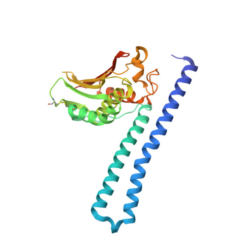
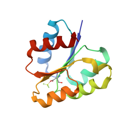
    Two-component signal transduction systems typically involve a sensor histidine kinase that specifically phosphorylates a single, cognate response regulator. This protein-protein interaction relies on molecular recognition via a small set of residues in each protein. To better understand how these residues determine the specificity of kinase-substrate interactions, we rationally rewired the interaction interface of a Thermotoga maritima two-component system, HK853-RR468, to match that found in a different two-component system, Escherichia coli PhoR-PhoB. The rewired proteins interacted robustly with each other, but no longer interacted with the parent proteins. Analysis of the crystal structures of the wild-type and mutant protein complexes and a systematic mutagenesis study reveal how individual mutations contribute to the rewiring of interaction specificity. Our approach and conclusions have implications for studies of other protein-protein interactions and protein evolution and for the design of novel protein interfaces.


  • Organizational Affiliation

    Computational and Systems Biology Initiative, Massachusetts Institute of Technology, Cambridge, MA 02139, USA; Department of Biology, Massachusetts Institute of Technology, Cambridge, MA 02139, USA.


Macromolecules
Find similar proteins by:  (by identity cutoff)  |  3D Structure
Entity ID: 1
MoleculeChains Sequence LengthOrganismDetailsImage
Histidine kinase
A, B
258Thermotoga maritima MSB8Mutation(s): 0 
Gene Names: TM_0853
EC: 2.7.13.3
UniProt
Find proteins for Q9WZV7 (Thermotoga maritima (strain ATCC 43589 / DSM 3109 / JCM 10099 / NBRC 100826 / MSB8))
Explore Q9WZV7 
Go to UniProtKB:  Q9WZV7
Entity Groups  
Sequence Clusters30% Identity50% Identity70% Identity90% Identity95% Identity100% Identity
UniProt GroupQ9WZV7
Sequence Annotations
Expand
  • Reference Sequence
Find similar proteins by:  (by identity cutoff)  |  3D Structure
Entity ID: 2
MoleculeChains Sequence LengthOrganismDetailsImage
Response regulator
C, D
122Thermotoga maritima MSB8Mutation(s): 4 
Gene Names: TM_0468
UniProt
Find proteins for Q9WYT9 (Thermotoga maritima (strain ATCC 43589 / DSM 3109 / JCM 10099 / NBRC 100826 / MSB8))
Explore Q9WYT9 
Go to UniProtKB:  Q9WYT9
Entity Groups  
Sequence Clusters30% Identity50% Identity70% Identity90% Identity95% Identity100% Identity
UniProt GroupQ9WYT9
Sequence Annotations
Expand
  • Reference Sequence
Small Molecules
Ligands 4 Unique
IDChains Name / Formula / InChI Key2D Diagram3D Interactions
ADP
Query on ADP

Download Ideal Coordinates CCD File 
H [auth A],
M [auth B]
ADENOSINE-5'-DIPHOSPHATE
C10 H15 N5 O10 P2
XTWYTFMLZFPYCI-KQYNXXCUSA-N
SO4
Query on SO4

Download Ideal Coordinates CCD File 
E [auth A],
F [auth A],
I [auth B],
J [auth B],
K [auth B]
SULFATE ION
O4 S
QAOWNCQODCNURD-UHFFFAOYSA-L
CL
Query on CL

Download Ideal Coordinates CCD File 
N [auth B]CHLORIDE ION
Cl
VEXZGXHMUGYJMC-UHFFFAOYSA-M
MG
Query on MG

Download Ideal Coordinates CCD File 
G [auth A],
L [auth B],
O [auth C],
P [auth D]
MAGNESIUM ION
Mg
JLVVSXFLKOJNIY-UHFFFAOYSA-N
Modified Residues  1 Unique
IDChains TypeFormula2D DiagramParent
BFD
Query on BFD
C, D
L-PEPTIDE LINKINGC4 H6 Be F3 N O4ASP
Experimental Data & Validation

Experimental Data

  • Method: X-RAY DIFFRACTION
  • Resolution: 3.10 Å
  • R-Value Free: 0.253 
  • R-Value Work: 0.202 
  • R-Value Observed: 0.204 
  • Space Group: C 2 2 21
Unit Cell:
Length ( Å )Angle ( ˚ )
a = 119.319α = 90
b = 143.928β = 90
c = 138.969γ = 90
Software Package:
Software NamePurpose
XDSdata scaling
PHASERphasing
REFMACrefinement
XDSdata reduction
SCALAdata scaling

Structure Validation

View Full Validation Report


Ligand Structure Quality Assessment 


Entry History 

Deposition Data

Revision History  (Full details and data files)

  • Version 1.0: 2013-09-04
    Type: Initial release
  • Version 1.1: 2013-09-25
    Changes: Database references, Structure summary
  • Version 1.2: 2023-11-08
    Changes: Data collection, Database references, Derived calculations, Refinement description