4J5T

Crystal structure of Processing alpha-Glucosidase I


Experimental Data Snapshot

  • Method: X-RAY DIFFRACTION
  • Resolution: 2.04 Å
  • R-Value Free: 0.235 
  • R-Value Work: 0.226 
  • R-Value Observed: 0.226 

wwPDB Validation   3D Report Full Report


This is version 2.0 of the entry. See complete history


Literature

Specificity of processing alpha-glucosidase I is guided by the substrate conformation: crystallographic and in silico studies.

Barker, M.K.Rose, D.R.

(2013) J Biol Chem 288: 13563-13574

  • DOI: https://doi.org/10.1074/jbc.M113.460436
  • Primary Citation of Related Structures:  
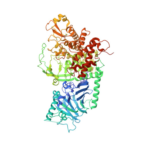
    4J5T

  • PubMed Abstract: 

    The enzyme “GluI” is key to the synthesis of critical glycoproteins in the cell. We have determined the structure of GluI, and modeled binding with its unique sugar substrate. The specificity of this interaction derives from a unique conformation of the substrate. Understanding the mechanism of the enzyme is of basic importance and relevant to potential development of antiviral inhibitors. Processing α-glucosidase I (GluI) is a key member of the eukaryotic N-glycosylation processing pathway, selectively catalyzing the first glycoprotein trimming step in the endoplasmic reticulum. Inhibition of GluI activity impacts the infectivity of enveloped viruses; however, despite interest in this protein from a structural, enzymatic, and therapeutic standpoint, little is known about its structure and enzymatic mechanism in catalysis of the unique glycan substrate Glc3Man9GlcNAc2. The first structural model of eukaryotic GluI is here presented at 2-Å resolution. Two catalytic residues are proposed, mutations of which result in catalytically inactive, properly folded protein. Using Autodocking methods with the known substrate and inhibitors as ligands, including a novel inhibitor characterized in this work, the active site of GluI was mapped. From these results, a model of substrate binding has been formulated, which is most likely conserved in mammalian GluI.


  • Organizational Affiliation

    Department of Medical Biophysics, University of Toronto, Ontario Cancer Institute, Princess Margaret Hospital, Toronto, Ontario M5G 2M9, Canada. megan.barker@utoronto.ca


Macromolecules
Find similar proteins by:  (by identity cutoff)  |  3D Structure
Entity ID: 1
MoleculeChains Sequence LengthOrganismDetailsImage
Mannosyl-oligosaccharide glucosidase811Saccharomyces cerevisiae S288CMutation(s): 0 
Gene Names: CWH41GLS1YGL027C
EC: 3.2.1.106
UniProt
Find proteins for P53008 (Saccharomyces cerevisiae (strain ATCC 204508 / S288c))
Explore P53008 
Go to UniProtKB:  P53008
Entity Groups  
Sequence Clusters30% Identity50% Identity70% Identity90% Identity95% Identity100% Identity
UniProt GroupP53008
Sequence Annotations
Expand
  • Reference Sequence
Oligosaccharides

Help

Entity ID: 2
MoleculeChains Length2D Diagram Glycosylation3D Interactions
2-acetamido-2-deoxy-beta-D-glucopyranose-(1-4)-2-acetamido-2-deoxy-beta-D-glucopyranose
B, C
2N-Glycosylation
Glycosylation Resources
GlyTouCan:  G42666HT
GlyCosmos:  G42666HT
GlyGen:  G42666HT
Experimental Data & Validation

Experimental Data

  • Method: X-RAY DIFFRACTION
  • Resolution: 2.04 Å
  • R-Value Free: 0.235 
  • R-Value Work: 0.226 
  • R-Value Observed: 0.226 
  • Space Group: P 21 21 21
Unit Cell:
Length ( Å )Angle ( ˚ )
a = 94.8α = 90
b = 101.8β = 90
c = 103.3γ = 90
Software Package:
Software NamePurpose
HKL-2000data collection
PHENIXmodel building
REFMACrefinement
HKL-2000data reduction
HKL-2000data scaling
PHENIXphasing

Structure Validation

View Full Validation Report


Entry History 

Deposition Data

Revision History  (Full details and data files)

  • Version 1.0: 2013-04-03
    Type: Initial release
  • Version 1.1: 2013-06-19
    Changes: Database references
  • Version 2.0: 2020-07-29
    Type: Remediation
    Reason: Carbohydrate remediation
    Changes: Advisory, Atomic model, Data collection, Database references, Derived calculations, Structure summary