Crystallization and preliminary X-ray diffraction studies of a surface mutant of the middle domain of PB2 from human influenza A (H1N1) virus
Tsurumura, T., Qiu, H., Yoshida, T., Tsumori, Y., Tsuge, H.(2014) Acta Crystallogr Sect F Struct Biol Cryst Commun 70: 72-75
- PubMed: 24419622
- DOI: https://doi.org/10.1107/S2053230X13032603
- Primary Citation of Related Structures:
4J2R - PubMed Abstract:
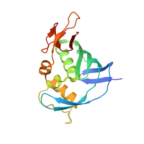
In the last hundred years, four influenza pandemics have been experienced, beginning with that in Spain in 1918. Influenza A virus causes severe pneumonia and its RNA polymerase is an important target for drug design. The influenza A (H1N1) virus has eight ribonucleoprotein complexes, which are composed of viral RNA, RNA polymerases and nucleoproteins. PB2 forms part of the RNA polymerase complex and plays an important role in binding to the cap structure of host mRNA. The middle domain of PB2 includes a cap-binding site. The structure of PB2 from H1N1 complexed with m(7)GTP has not been reported. Plate-like crystals of the middle domain of PB2 from H1N1 were obtained, but the quality of these crystals was not good. An attempt was made to crystallize the middle domain of PB2 complexed with m(7)GTP using a soaking method; however, electron density for m(7)GTP was not observed on preliminary X-ray diffraction analysis. This protein has hydrophobic residues on its surface and is stable in the presence of high salt concentrations. To improve the solubility, a surface double mutant (P453H and I471T) was prepared. These mutations change the surface electrostatic potential drastically. The protein was successfully prepared at a lower salt concentration and good cube-shaped crystals were obtained using this protein. Here, the crystallization and preliminary X-ray diffraction analysis of this mutant of the middle domain of PB2 are reported.
Organizational Affiliation:
Faculty of Life Sciences, Kyoto Sangyo University, Kamigamo-Motoyama, Kyoto 603-8555, Japan.


