4IUF

Crystal Structure of Human TDP-43 RRM1 Domain in Complex with a Single-stranded DNA


Experimental Data Snapshot

  • Method: X-RAY DIFFRACTION
  • Resolution: 2.75 Å
  • R-Value Free: 0.259 
  • R-Value Work: 0.206 
  • R-Value Observed: 0.212 

wwPDB Validation   3D Report Full Report


This is version 1.2 of the entry. See complete history


Literature

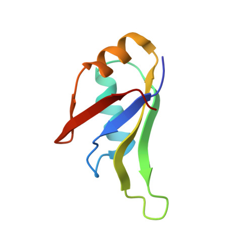
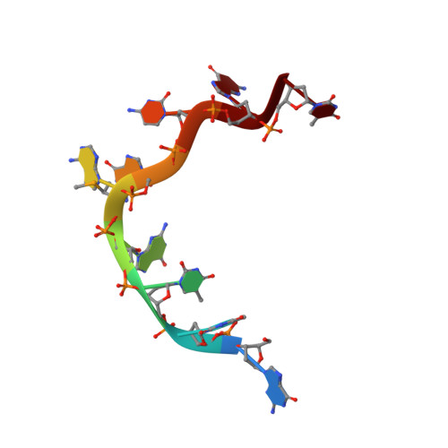
The crystal structure of TDP-43 RRM1-DNA complex reveals the specific recognition for UG- and TG-rich nucleic acids.

Kuo, P.H.Chiang, C.H.Wang, Y.T.Doudeva, L.G.Yuan, H.S.

(2014) Nucleic Acids Res 42: 4712-4722

  • DOI: https://doi.org/10.1093/nar/gkt1407
  • Primary Citation of Related Structures:  
    4IUF

  • PubMed Abstract: 

    TDP-43 is an important pathological protein that aggregates in the diseased neuronal cells and is linked to various neurodegenerative disorders. In normal cells, TDP-43 is primarily an RNA-binding protein; however, how the dimeric TDP-43 binds RNA via its two RNA recognition motifs, RRM1 and RRM2, is not clear. Here we report the crystal structure of human TDP-43 RRM1 in complex with a single-stranded DNA showing that RRM1 binds the nucleic acid extensively not only by the conserved β-sheet residues but also by the loop residues. Mutational and biochemical assays further reveal that both RRMs in TDP-43 dimers participate in binding of UG-rich RNA or TG-rich DNA with RRM1 playing a dominant role and RRM2 playing a supporting role. Moreover, RRM1 of the amyotrophic lateral sclerosis-linked mutant D169G binds DNA as efficiently as the wild type; nevertheless, it is more resistant to thermal denaturation, suggesting that the resistance to degradation is likely linked to TDP-43 proteinopathies. Taken together all the data, we suggest a model showing that the two RRMs in each protomer of TDP-43 homodimer work together in RNA binding and thus the dimeric TDP-43 recognizes long clusters of UG-rich RNA to achieve high affinity and specificity.


  • Organizational Affiliation

    Institute of Molecular Biology, Academia Sinica, Taipei, Taiwan, Institute of Bioinformatics and Structural Biology, National Tsing Hua University, Hsin Chu, Taiwan and Graduate Institute of Biochemistry and Molecular Biology, National Taiwan University, Taipei 10048, Taiwan.


Macromolecules

Find similar proteins by:  (by identity cutoff)  |  3D Structure
Entity ID: 1
MoleculeChains Sequence LengthOrganismDetailsImage
TAR DNA-binding protein 4377Homo sapiensMutation(s): 0 
Gene Names: TARDBPTDP43
UniProt & NIH Common Fund Data Resources
Find proteins for Q13148 (Homo sapiens)
Explore Q13148 
Go to UniProtKB:  Q13148
PHAROS:  Q13148
GTEx:  ENSG00000120948 
Entity Groups  
Sequence Clusters30% Identity50% Identity70% Identity90% Identity95% Identity100% Identity
UniProt GroupQ13148
Sequence Annotations
Expand
  • Reference Sequence

Find similar nucleic acids by:  Sequence   |   3D Structure  

Entity ID: 2
MoleculeChains LengthOrganismImage
5'-D(*GP*TP*TP*GP*(XUA)P*GP*CP*GP*T)-3'9N/A
Sequence Annotations
Expand
  • Reference Sequence
Experimental Data & Validation

Experimental Data

  • Method: X-RAY DIFFRACTION
  • Resolution: 2.75 Å
  • R-Value Free: 0.259 
  • R-Value Work: 0.206 
  • R-Value Observed: 0.212 
  • Space Group: P 65 2 2
Unit Cell:
Length ( Å )Angle ( ˚ )
a = 71.11α = 90
b = 71.11β = 90
c = 101.663γ = 120
Software Package:
Software NamePurpose
PHENIXrefinement
PDB_EXTRACTdata extraction
HKL-2000data collection
HKL-2000data reduction
HKL-2000data scaling
PHENIXphasing

Structure Validation

View Full Validation Report


Entry History 

Deposition Data

Revision History  (Full details and data files)

  • Version 1.0: 2014-01-29
    Type: Initial release
  • Version 1.1: 2014-10-08
    Changes: Database references
  • Version 1.2: 2024-02-28
    Changes: Data collection, Database references, Derived calculations