Increasing the structural coverage of tuberculosis drug targets.
Baugh, L., Phan, I., Begley, D.W., Clifton, M.C., Armour, B., Dranow, D.M., Taylor, B.M., Muruthi, M.M., Abendroth, J., Fairman, J.W., Fox, D., Dieterich, S.H., Staker, B.L., Gardberg, A.S., Choi, R., Hewitt, S.N., Napuli, A.J., Myers, J., Barrett, L.K., Zhang, Y., Ferrell, M., Mundt, E., Thompkins, K., Tran, N., Lyons-Abbott, S., Abramov, A., Sekar, A., Serbzhinskiy, D., Lorimer, D., Buchko, G.W., Stacy, R., Stewart, L.J., Edwards, T.E., Van Voorhis, W.C., Myler, P.J.(2015) Tuberculosis (Edinb) 95: 142-148
- PubMed: 25613812
- DOI: https://doi.org/10.1016/j.tube.2014.12.003
- Primary Citation of Related Structures:
3GVC, 3GVG, 3GWC, 3H7F, 3H81, 3HE2, 3HWI, 3HWK, 3HZG, 3ICO, 3KHP, 3LLS, 3MOY, 3MPZ, 3MYB, 3NDN, 3NDO, 3NF4, 3NG3, 3NJD, 3NWO, 3O0M, 3O38, 3OC6, 3OC7, 3OI9, 3OKS, 3OME, 3P0T, 3P2Y, 3P4I, 3P4T, 3P5M, 3P85, 3PE8, 3PK0, 3PPI, 3PZY, 3Q1T, 3Q8N, 3QBP, 3QDF, 3QHA, 3QIV, 3QK8, 3QKA, 3QLJ, 3QMJ, 3QRE, 3QUA - PubMed Abstract:
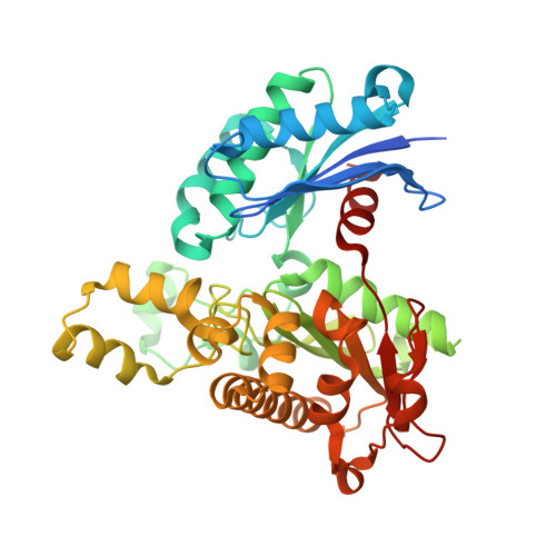
High-resolution three-dimensional structures of essential Mycobacterium tuberculosis (Mtb) proteins provide templates for TB drug design, but are available for only a small fraction of the Mtb proteome. Here we evaluate an intra-genus "homolog-rescue" strategy to increase the structural information available for TB drug discovery by using mycobacterial homologs with conserved active sites. Of 179 potential TB drug targets selected for x-ray structure determination, only 16 yielded a crystal structure. By adding 1675 homologs from nine other mycobacterial species to the pipeline, structures representing an additional 52 otherwise intractable targets were solved. To determine whether these homolog structures would be useful surrogates in TB drug design, we compared the active sites of 106 pairs of Mtb and non-TB mycobacterial (NTM) enzyme homologs with experimentally determined structures, using three metrics of active site similarity, including superposition of continuous pharmacophoric property distributions. Pair-wise structural comparisons revealed that 19/22 pairs with >55% overall sequence identity had active site Cα RMSD <1 Å, >85% side chain identity, and ≥80% PSAPF (similarity based on pharmacophoric properties) indicating highly conserved active site shape and chemistry. Applying these results to the 52 NTM structures described above, 41 shared >55% sequence identity with the Mtb target, thus increasing the effective structural coverage of the 179 Mtb targets over three-fold (from 9% to 32%). The utility of these structures in TB drug design can be tested by designing inhibitors using the homolog structure and assaying the cognate Mtb enzyme; a promising test case, Mtb cytidylate kinase, is described. The homolog-rescue strategy evaluated here for TB is also generalizable to drug targets for other diseases.
Organizational Affiliation:
Seattle Structural Genomics Center for Infectious Disease, United States; Seattle Biomedical Research Institute, 307 Westlake Ave N, Suite 500, Seattle, WA 98109, United States.