Crystal and Solution Studies of the "Plus-C" Odorant-binding Protein 48 from Anopheles gambiae: CONTROL OF BINDING SPECIFICITY THROUGH THREE-DIMENSIONAL DOMAIN SWAPPING.
Tsitsanou, K.E., Drakou, C.E., Thireou, T., Vitlin Gruber, A., Kythreoti, G., Azem, A., Fessas, D., Eliopoulos, E., Iatrou, K., Zographos, S.E.(2013) J Biol Chem 288: 33427-33438
- PubMed: 24097978
- DOI: https://doi.org/10.1074/jbc.M113.505289
- Primary Citation of Related Structures:
4IJ7, 4KYN - PubMed Abstract:
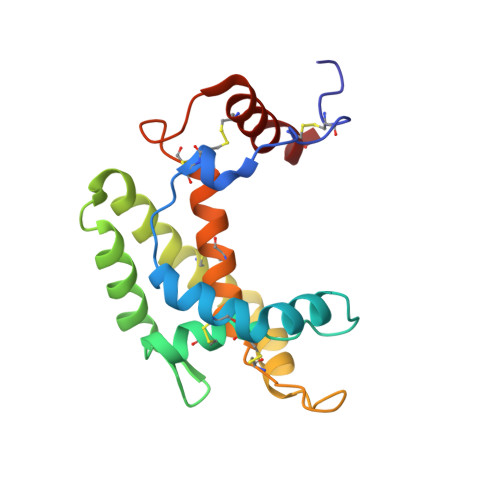
Much physiological and behavioral evidence has been provided suggesting that insect odorant-binding proteins (OBPs) are indispensable for odorant recognition and thus are appealing targets for structure-based discovery and design of novel host-seeking disruptors. Despite the fact that more than 60 putative OBP-encoding genes have been identified in the malaria vector Anopheles gambiae, the crystal structures of only six of them are known. It is therefore clear that OBP structure determination constitutes the bottleneck for structure-based approaches to mosquito repellent/attractant discovery. Here, we describe the three-dimensional structure of an A. gambiae "Plus-C" group OBP (AgamOBP48), which exhibits the second highest expression levels in female antennae. This structure represents the first example of a three-dimensional domain-swapped dimer in dipteran species. A combined binding site is formed at the dimer interface by equal contribution of each monomer. Structural comparisons with the monomeric AgamOBP47 revealed that the major structural difference between the two Plus-C proteins localizes in their N- and C-terminal regions, and their concerted conformational change may account for monomer-swapped dimer conversion and furthermore the formation of novel binding pockets. Using a combination of gel filtration chromatography, differential scanning calorimetry, and analytical ultracentrifugation, we demonstrate the AgamOBP48 dimerization in solution. Eventually, molecular modeling calculations were used to predict the binding mode of the most potent synthetic ligand of AgamOBP48 known so far, discovered by ligand- and structure-based virtual screening. The structure-aided identification of multiple OBP binders represents a powerful tool to be employed in the effort to control transmission of the vector-borne diseases.
Organizational Affiliation:
From the Institute of Biology, Medicinal Chemistry and Biotechnology, National Hellenic Research Foundation, 48 Vassileos Constantinou Avenue, 11635 Athens, Greece.