4IGL

Structure of the RHS-repeat containing BC component of the secreted ABC toxin complex from Yersinia entomophaga


Experimental Data Snapshot

  • Method: X-RAY DIFFRACTION
  • Resolution: 2.49 Å
  • R-Value Free: 0.251 
  • R-Value Work: 0.200 
  • R-Value Observed: 0.203 

wwPDB Validation   3D Report Full Report


This is version 1.3 of the entry. See complete history


Literature

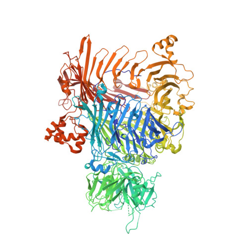
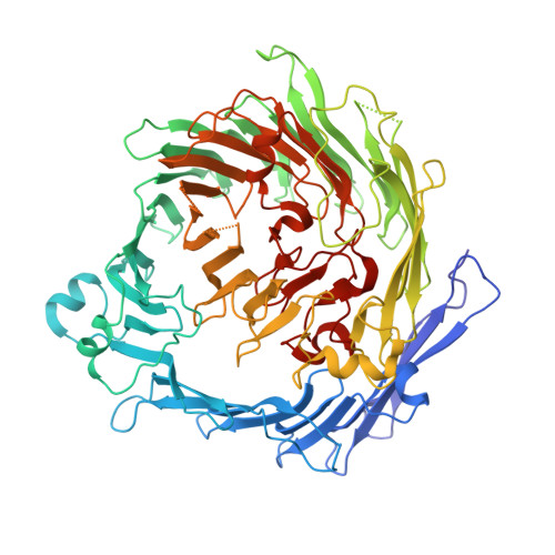
The BC component of ABC toxins is an RHS-repeat-containing protein encapsulation device

Busby, J.N.Panjikar, S.Landsberg, M.J.Hurst, M.R.H.Lott, J.S.

(2013) Nature 501: 547-550

  • DOI: https://doi.org/10.1038/nature12465
  • Primary Citation of Related Structures:  
    4DWS, 4IGL

  • PubMed Abstract: 

    The ABC toxin complexes produced by certain bacteria are of interest owing to their potent insecticidal activity and potential role in human disease. These complexes comprise at least three proteins (A, B and C), which must assemble to be fully toxic. The carboxy-terminal region of the C protein is the main cytotoxic component, and is poorly conserved between different toxin complexes. A general model of action has been proposed, in which the toxin complex binds to the cell surface via the A protein, is endocytosed, and subsequently forms a pH-triggered channel, allowing the translocation of C into the cytoplasm, where it can cause cytoskeletal disruption in both insect and mammalian cells. Toxin complexes have been visualized using single-particle electron microscopy, but no high-resolution structures of the components are available, and the role of the B protein in the mechanism of toxicity remains unknown. Here we report the three-dimensional structure of the complex formed between the B and C proteins, determined to 2.5 Å by X-ray crystallography. These proteins assemble to form an unprecedented, large hollow structure that encapsulates and sequesters the cytotoxic, C-terminal region of the C protein like the shell of an egg. The shell is decorated on one end by a β-propeller domain, which mediates attachment of the B-C heterodimer to the A protein in the native complex. The structure reveals how C auto-proteolyses when folded in complex with B. The C protein is the first example, to our knowledge, of a structure that contains rearrangement hotspot (RHS) repeats, and illustrates a marked structural architecture that is probably conserved across both this widely distributed bacterial protein family and the related eukaryotic tyrosine-aspartate (YD)-repeat-containing protein family, which includes the teneurins. The structure provides the first clues about the function of these protein repeat families, and suggests a generic mechanism for protein encapsulation and delivery.


  • Organizational Affiliation

    AgResearch Structural Biology Laboratory, School of Biological Sciences, The University of Auckland, Auckland 1142, New Zealand.


Macromolecules
Find similar proteins by:  (by identity cutoff)  |  3D Structure
Entity ID: 1
MoleculeChains Sequence LengthOrganismDetailsImage
YenB
A, C
1,488Yersinia entomophagaMutation(s): 0 
Gene Names: yenB
UniProt
Find proteins for B6A880 (Yersinia entomophaga)
Explore B6A880 
Go to UniProtKB:  B6A880
Entity Groups  
Sequence Clusters30% Identity50% Identity70% Identity90% Identity95% Identity100% Identity
UniProt GroupB6A880
Sequence Annotations
Expand
  • Reference Sequence
Find similar proteins by:  (by identity cutoff)  |  3D Structure
Entity ID: 2
MoleculeChains Sequence LengthOrganismDetailsImage
YenC2
B, D
690Yersinia entomophagaMutation(s): 0 
Gene Names: yenC2
UniProt
Find proteins for B6A882 (Yersinia entomophaga)
Explore B6A882 
Go to UniProtKB:  B6A882
Entity Groups  
Sequence Clusters30% Identity50% Identity70% Identity90% Identity95% Identity100% Identity
UniProt GroupB6A882
Sequence Annotations
Expand
  • Reference Sequence
Small Molecules
Ligands 2 Unique
IDChains Name / Formula / InChI Key2D Diagram3D Interactions
GOL
Query on GOL

Download Ideal Coordinates CCD File 
E [auth A]
F [auth A]
J [auth C]
K [auth C]
L [auth C]
E [auth A],
F [auth A],
J [auth C],
K [auth C],
L [auth C],
M [auth C],
N [auth C],
R [auth D]
GLYCEROL
C3 H8 O3
PEDCQBHIVMGVHV-UHFFFAOYSA-N
K
Query on K

Download Ideal Coordinates CCD File 
G [auth A]
H [auth A]
I [auth A]
O [auth C]
P [auth C]
G [auth A],
H [auth A],
I [auth A],
O [auth C],
P [auth C],
Q [auth C]
POTASSIUM ION
K
NPYPAHLBTDXSSS-UHFFFAOYSA-N
Experimental Data & Validation

Experimental Data

  • Method: X-RAY DIFFRACTION
  • Resolution: 2.49 Å
  • R-Value Free: 0.251 
  • R-Value Work: 0.200 
  • R-Value Observed: 0.203 
  • Space Group: P 21 21 21
Unit Cell:
Length ( Å )Angle ( ˚ )
a = 133.719α = 90
b = 147.596β = 90
c = 274.354γ = 90
Software Package:
Software NamePurpose
Aimlessdata scaling
PHASERphasing
PHENIXrefinement
PDB_EXTRACTdata extraction
Blu-Icedata collection
XDSdata reduction

Structure Validation

View Full Validation Report


Entry History 

Deposition Data

Revision History  (Full details and data files)

  • Version 1.0: 2013-08-07
    Type: Initial release
  • Version 1.1: 2013-10-02
    Changes: Database references
  • Version 1.2: 2024-03-20
    Changes: Data collection, Database references, Derived calculations
  • Version 1.3: 2024-04-03
    Changes: Refinement description