4ICA

Crystal structure of a C-terminal truncated form of the matrix subunit (p15) of Feline Immunodeficiency Virus (FIV)


Experimental Data Snapshot

  • Method: X-RAY DIFFRACTION
  • Resolution: 2.70 Å
  • R-Value Free: 0.279 
  • R-Value Work: 0.212 
  • R-Value Observed: 0.215 

wwPDB Validation   3D Report Full Report


This is version 1.2 of the entry. See complete history


Literature

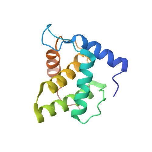
Biophysical characterization and crystal structure of the Feline Immunodeficiency Virus p15 matrix protein.

Serriere, J.Robert, X.Perez, M.Gouet, P.Guillon, C.

(2013) Retrovirology 10: 64-64

  • DOI: https://doi.org/10.1186/1742-4690-10-64
  • Primary Citation of Related Structures:  
    4IC9, 4ICA

  • PubMed Abstract: 

    Feline Immunodeficiency Virus (FIV) is a viral pathogen that infects domestic cats and wild felids. During the viral replication cycle, the FIV p15 matrix protein oligomerizes to form a closed matrix that underlies the lipidic envelope of the virion. Because of its crucial role in the early and late stages of viral morphogenesis, especially in viral assembly, FIV p15 is an interesting target in the development of potential new therapeutic strategies.


Macromolecules
Find similar proteins by:  (by identity cutoff)  |  3D Structure
Entity ID: 1
MoleculeChains Sequence LengthOrganismDetailsImage
Matrix protein p15
A, B
134Feline immunodeficiency virus (isolate Petaluma)Mutation(s): 0 
Gene Names: FIV Gaggag
UniProt
Find proteins for P16087 (Feline immunodeficiency virus (isolate Petaluma))
Explore P16087 
Go to UniProtKB:  P16087
Entity Groups  
Sequence Clusters30% Identity50% Identity70% Identity90% Identity95% Identity100% Identity
UniProt GroupP16087
Sequence Annotations
Expand
  • Reference Sequence
Small Molecules
Ligands 1 Unique
IDChains Name / Formula / InChI Key2D Diagram3D Interactions
7PE
Query on 7PE

Download Ideal Coordinates CCD File 
C [auth A],
D [auth B]
2-(2-(2-(2-(2-(2-ETHOXYETHOXY)ETHOXY)ETHOXY)ETHOXY)ETHOXY)ETHANOL
C14 H30 O7
UKXKPKBTMYNOFS-UHFFFAOYSA-N
Experimental Data & Validation

Experimental Data

  • Method: X-RAY DIFFRACTION
  • Resolution: 2.70 Å
  • R-Value Free: 0.279 
  • R-Value Work: 0.212 
  • R-Value Observed: 0.215 
  • Space Group: P 1 21 1
Unit Cell:
Length ( Å )Angle ( ˚ )
a = 28.48α = 90
b = 71.3β = 99.63
c = 57.06γ = 90
Software Package:
Software NamePurpose
MxCuBEdata collection
MrBUMPphasing
PHENIXrefinement
XDSdata reduction
XSCALEdata scaling

Structure Validation

View Full Validation Report


Entry History 

Deposition Data

Revision History  (Full details and data files)

  • Version 1.0: 2013-07-10
    Type: Initial release
  • Version 1.1: 2024-02-28
    Changes: Data collection, Database references, Derived calculations
  • Version 1.2: 2024-04-03
    Changes: Refinement description