4I5I

Crystal structure of the SIRT1 catalytic domain bound to NAD and an EX527 analog


Experimental Data Snapshot

  • Method: X-RAY DIFFRACTION
  • Resolution: 2.50 Å
  • R-Value Free: 0.195 
  • R-Value Work: 0.167 
  • R-Value Observed: 0.169 

wwPDB Validation   3D Report Full Report


Ligand Structure Quality Assessment 


This is version 1.3 of the entry. See complete history


Literature

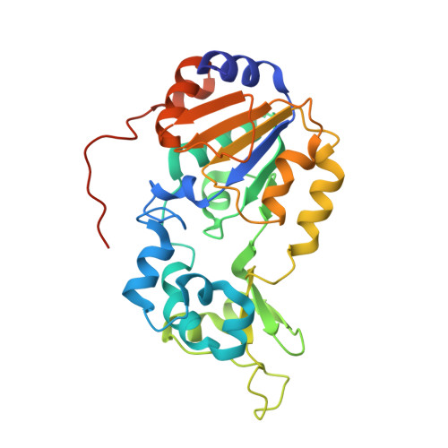
The 2.5 angstrom crystal structure of the SIRT1 catalytic domain bound to nicotinamide adenine dinucleotide (NAD+) and an indole (EX527 analogue) reveals a novel mechanism of histone deacetylase inhibition.

Zhao, X.Allison, D.Condon, B.Zhang, F.Gheyi, T.Zhang, A.Ashok, S.Russell, M.MacEwan, I.Qian, Y.Jamison, J.A.Luz, J.G.

(2013) J Med Chem 56: 963-969

  • DOI: https://doi.org/10.1021/jm301431y
  • Primary Citation of Related Structures:  
    4I5I

  • PubMed Abstract: 

    The sirtuin SIRT1 is a NAD(+)-dependent histone deacetylase, a Sir2 family member, and one of seven human sirtuins. Sirtuins are conserved from archaea to mammals and regulate transcription, genome stability, longevity, and metabolism. SIRT1 regulates transcription via deacetylation of transcription factors such as PPARγ, NFκB, and the tumor suppressor protein p53. EX527 (27) is a nanomolar SIRT1 inhibitor and a micromolar SIRT2 inhibitor. To elucidate the mechanism of SIRT inhibition by 27, we determined the 2.5 Å crystal structure of the SIRT1 catalytic domain (residues 241-516) bound to NAD(+) and the 27 analogue compound 35. 35 binds deep in the catalytic cleft, displacing the NAD(+) nicotinamide and forcing the cofactor into an extended conformation. The extended NAD(+) conformation sterically prevents substrate binding. The SIRT1/NAD(+)/35 crystal structure defines a novel mechanism of histone deacetylase inhibition and provides a basis for understanding, and rationally improving, inhibition of this therapeutically important target by drug-like molecules.


  • Organizational Affiliation

    Lilly Biotechnology Center San Diego, 10300 Campus Point Drive, Suite 200, San Diego, California 92121, USA.


Macromolecules
Find similar proteins by:  (by identity cutoff)  |  3D Structure
Entity ID: 1
MoleculeChains Sequence LengthOrganismDetailsImage
NAD-dependent protein deacetylase sirtuin-1
A, B
287Homo sapiensMutation(s): 0 
Gene Names: SIRT1SIR2L1
EC: 3.5.1
UniProt & NIH Common Fund Data Resources
Find proteins for Q96EB6 (Homo sapiens)
Explore Q96EB6 
Go to UniProtKB:  Q96EB6
PHAROS:  Q96EB6
GTEx:  ENSG00000096717 
Entity Groups  
Sequence Clusters30% Identity50% Identity70% Identity90% Identity95% Identity100% Identity
UniProt GroupQ96EB6
Sequence Annotations
Expand
  • Reference Sequence
Experimental Data & Validation

Experimental Data

  • Method: X-RAY DIFFRACTION
  • Resolution: 2.50 Å
  • R-Value Free: 0.195 
  • R-Value Work: 0.167 
  • R-Value Observed: 0.169 
  • Space Group: P 32
Unit Cell:
Length ( Å )Angle ( ˚ )
a = 106.513α = 90
b = 106.513β = 90
c = 80.663γ = 120
Software Package:
Software NamePurpose
PHASERphasing
REFMACrefinement
SCALAdata scaling

Structure Validation

View Full Validation Report


Ligand Structure Quality Assessment 


Entry History 

Deposition Data

Revision History  (Full details and data files)

  • Version 1.0: 2013-01-23
    Type: Initial release
  • Version 1.1: 2013-08-28
    Changes: Database references
  • Version 1.2: 2014-11-12
    Changes: Structure summary
  • Version 1.3: 2023-09-20
    Changes: Data collection, Database references, Derived calculations, Refinement description