4I1S

Melanoma differentiation associated protein-5 Helicase domain complex with inhibitor Non-structural protein V


Experimental Data Snapshot

  • Method: X-RAY DIFFRACTION
  • Resolution: 2.29 Å
  • R-Value Free: 0.230 
  • R-Value Work: 0.179 
  • R-Value Observed: 0.182 

wwPDB Validation   3D Report Full Report


This is version 1.2 of the entry. See complete history


Literature

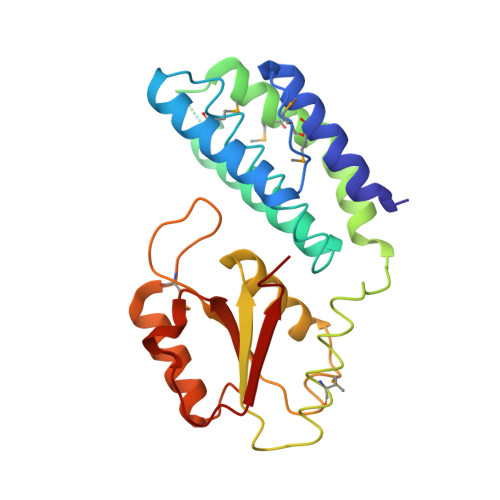
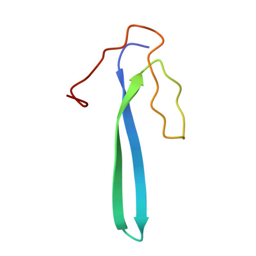
Paramyxovirus V proteins disrupt the fold of the RNA sensor MDA5 to inhibit antiviral signaling.

Motz, C.Schuhmann, K.M.Kirchhofer, A.Moldt, M.Witte, G.Conzelmann, K.K.Hopfner, K.P.

(2013) Science 339: 690-693

  • DOI: https://doi.org/10.1126/science.1230949
  • Primary Citation of Related Structures:  
    4I1S

  • PubMed Abstract: 

    The retinoic acid-inducible gene I (RIG-I)-like receptor (RLR) melanoma differentiation-associated protein 5 (MDA5) senses cytoplasmic viral RNA and activates antiviral innate immunity. To reveal how paramyxoviruses counteract this response, we determined the crystal structure of the MDA5 adenosine 5'-triphosphate (ATP)-hydrolysis domain in complex with the viral inhibitor V protein. The V protein unfolded the ATP-hydrolysis domain of MDA5 via a β-hairpin motif and recognized a structural motif of MDA5 that is normally buried in the conserved helicase fold. This leads to disruption of the MDA5 ATP-hydrolysis site and prevention of RNA-bound MDA5 filament formation. The structure explains why V proteins inactivate MDA5, but not RIG-I, and mutating only two amino acids in RIG-I induces robust V protein binding. Our results suggest an inhibition mechanism of RLR signalosome formation by unfolding of receptor and inhibitor.


  • Organizational Affiliation

    Department of Biochemistry and Gene Center, Ludwig-Maximilians-University, Munich, Germany.


Macromolecules
Find similar proteins by:  (by identity cutoff)  |  3D Structure
Entity ID: 1
MoleculeChains Sequence LengthOrganismDetailsImage
Melanoma differentiation associated protein-5243Sus scrofaMutation(s): 0 
Gene Names: MDA5
UniProt
Find proteins for A7LCX1 (Sus scrofa)
Explore A7LCX1 
Go to UniProtKB:  A7LCX1
Entity Groups  
Sequence Clusters30% Identity50% Identity70% Identity90% Identity95% Identity100% Identity
UniProt GroupA7LCX1
Sequence Annotations
Expand
  • Reference Sequence
Find similar proteins by:  (by identity cutoff)  |  3D Structure
Entity ID: 2
MoleculeChains Sequence LengthOrganismDetailsImage
Non-structural protein V52Simian virus 5 (strain W3)Mutation(s): 0 
Gene Names: P/V
UniProt
Find proteins for P11207 (Parainfluenza virus 5 (strain W3))
Explore P11207 
Go to UniProtKB:  P11207
Entity Groups  
Sequence Clusters30% Identity50% Identity70% Identity90% Identity95% Identity100% Identity
UniProt GroupP11207
Sequence Annotations
Expand
  • Reference Sequence
Small Molecules
Modified Residues  1 Unique
IDChains TypeFormula2D DiagramParent
MSE
Query on MSE
A
L-PEPTIDE LINKINGC5 H11 N O2 SeMET
Experimental Data & Validation

Experimental Data

  • Method: X-RAY DIFFRACTION
  • Resolution: 2.29 Å
  • R-Value Free: 0.230 
  • R-Value Work: 0.179 
  • R-Value Observed: 0.182 
  • Space Group: P 21 21 21
Unit Cell:
Length ( Å )Angle ( ˚ )
a = 51.07α = 90
b = 52.49β = 90
c = 120.79γ = 90
Software Package:
Software NamePurpose
DNAdata collection
SHARPphasing
PHENIXrefinement
XDSdata reduction
XSCALEdata scaling

Structure Validation

View Full Validation Report


Entry History 

Deposition Data

Revision History  (Full details and data files)

  • Version 1.0: 2013-01-30
    Type: Initial release
  • Version 1.1: 2013-02-20
    Changes: Database references
  • Version 1.2: 2017-07-26
    Changes: Advisory, Refinement description, Source and taxonomy