4HJO

Crystal structure of the inactive EGFR tyrosine kinase domain with erlotinib


Experimental Data Snapshot

  • Method: X-RAY DIFFRACTION
  • Resolution: 2.75 Å
  • R-Value Free: 0.253 
  • R-Value Work: 0.230 
  • R-Value Observed: 0.231 

wwPDB Validation   3D Report Full Report


Ligand Structure Quality Assessment 


This is version 1.2 of the entry. See complete history


Literature

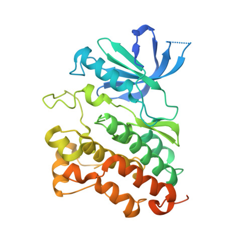
Erlotinib binds both inactive and active conformations of the EGFR tyrosine kinase domain.

Park, J.H.Liu, Y.Lemmon, M.A.Radhakrishnan, R.

(2012) Biochem J 448: 417-423

  • DOI: https://doi.org/10.1042/BJ20121513
  • Primary Citation of Related Structures:  
    4HJO

  • PubMed Abstract: 

    Erlotinib and gefitinib, tyrosine kinase inhibitors used to block EGFR (epidermal growth factor receptor) signalling in cancer, are thought to bind only the active conformation of the EGFR-TKD (tyrosine kinase domain). Through parallel computational and crystallographic studies, we show in the present study that erlotinib also binds the inactive EGFR-TKD conformation, which may have significant implications for its use in EGFR-mutated cancers.


  • Organizational Affiliation

    Department of Biochemistry and Biophysics, Perelman School of Medicine, University of Pennsylvania, Philadelphia, PA 19104, USA.


Macromolecules
Find similar proteins by:  (by identity cutoff)  |  3D Structure
Entity ID: 1
MoleculeChains Sequence LengthOrganismDetailsImage
Epidermal growth factor receptor337Homo sapiensMutation(s): 1 
Gene Names: EGFRERBBERBB1HER1
EC: 2.7.10.1
UniProt & NIH Common Fund Data Resources
Find proteins for P00533 (Homo sapiens)
Explore P00533 
Go to UniProtKB:  P00533
PHAROS:  P00533
GTEx:  ENSG00000146648 
Entity Groups  
Sequence Clusters30% Identity50% Identity70% Identity90% Identity95% Identity100% Identity
UniProt GroupP00533
Sequence Annotations
Expand
  • Reference Sequence
Small Molecules
Ligands 1 Unique
IDChains Name / Formula / InChI Key2D Diagram3D Interactions
AQ4
Query on AQ4

Download Ideal Coordinates CCD File 
B [auth A][6,7-BIS(2-METHOXY-ETHOXY)QUINAZOLINE-4-YL]-(3-ETHYNYLPHENYL)AMINE
C22 H23 N3 O4
AAKJLRGGTJKAMG-UHFFFAOYSA-N
Binding Affinity Annotations 
IDSourceBinding Affinity
AQ4 BindingDB:  4HJO Ki: min: 0.1, max: 136 (nM) from 6 assay(s)
Kd: min: 0.35, max: 970 (nM) from 15 assay(s)
IC50: min: 0.03, max: 260 (nM) from 53 assay(s)
Experimental Data & Validation

Experimental Data

  • Method: X-RAY DIFFRACTION
  • Resolution: 2.75 Å
  • R-Value Free: 0.253 
  • R-Value Work: 0.230 
  • R-Value Observed: 0.231 
  • Space Group: C 2 2 21
Unit Cell:
Length ( Å )Angle ( ˚ )
a = 77.967α = 90
b = 114.284β = 90
c = 84.918γ = 90
Software Package:
Software NamePurpose
HKL-2000data collection
PHASERphasing
REFMACrefinement
HKL-2000data reduction
HKL-2000data scaling

Structure Validation

View Full Validation Report


Ligand Structure Quality Assessment 


Entry History 

Deposition Data

Revision History  (Full details and data files)

  • Version 1.0: 2012-11-14
    Type: Initial release
  • Version 1.1: 2012-12-05
    Changes: Database references
  • Version 1.2: 2023-09-20
    Changes: Data collection, Database references, Derived calculations, Refinement description