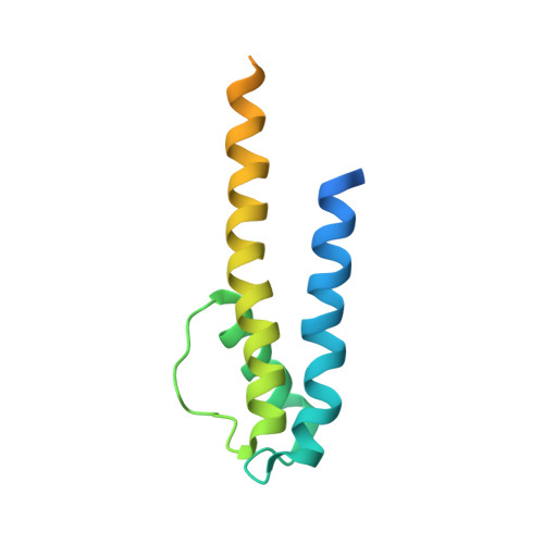
4H37

Crystal structure of a voltage-gated K+ channel pore domain in a closed state in lipid membranes


Experimental Data Snapshot

  • Method: X-RAY DIFFRACTION
  • Resolution: 3.35 Å
  • R-Value Free: 0.341 
  • R-Value Work: 0.314 
  • R-Value Observed: 0.318 

wwPDB Validation   3D Report Full Report


This is version 1.3 of the entry. See complete history


Literature

Crystal Structure of a Voltage-gated K+ Channel Pore Module in a Closed State in Lipid Membranes.

Santos, J.S.Asmar-Rovira, G.A.Han, G.W.Liu, W.Syeda, R.Cherezov, V.Baker, K.A.Stevens, R.C.Montal, M.

(2012) J Biol Chem 287: 43063-43070

  • DOI: https://doi.org/10.1074/jbc.M112.415091
  • Primary Citation of Related Structures:  
    4H33, 4H37

  • PubMed Abstract: 

    Voltage-gated K(+) channels underlie the electrical excitability of cells. Each subunit of the functional tetramer consists of the tandem fusion of two modules, an N-terminal voltage-sensor and a C-terminal pore. To investigate how sensor coupling to the pore generates voltage-dependent channel opening, we solved the crystal structure and characterized the function of a voltage-gated K(+) channel pore in a lipid membrane. The structure of a functional channel in a membrane environment at 3.1 Å resolution establishes an unprecedented connection between channel structure and function. The structure is unique in delineating an ion-occupied ready to conduct selectivity filter, a confined aqueous cavity, and a closed activation gate, embodying a dynamic entity trapped in an unstable closed state.


  • Organizational Affiliation

    Section of Neurobiology, Division of Biological Sciences, University of California San Diego, La Jolla, California 92093, USA.


Macromolecules
Find similar proteins by:  (by identity cutoff)  |  3D Structure
Entity ID: 1
MoleculeChains Sequence LengthOrganismDetailsImage
Lmo2059 protein
A, B
137Listeria monocytogenes EGD-eMutation(s): 1 
Gene Names: lmo2059
Membrane Entity: Yes 
UniProt
Find proteins for Q8Y5K1 (Listeria monocytogenes serovar 1/2a (strain ATCC BAA-679 / EGD-e))
Explore Q8Y5K1 
Go to UniProtKB:  Q8Y5K1
Entity Groups  
Sequence Clusters30% Identity50% Identity70% Identity90% Identity95% Identity100% Identity
UniProt GroupQ8Y5K1
Sequence Annotations
Expand
  • Reference Sequence
Experimental Data & Validation

Experimental Data

  • Method: X-RAY DIFFRACTION
  • Resolution: 3.35 Å
  • R-Value Free: 0.341 
  • R-Value Work: 0.314 
  • R-Value Observed: 0.318 
  • Space Group: P 21 21 2
Unit Cell:
Length ( Å )Angle ( ˚ )
a = 56.1α = 90
b = 80.67β = 90
c = 55.522γ = 90
Software Package:
Software NamePurpose
Blu-Icedata collection
PHASERphasing
BUSTERrefinement
HKL-2000data reduction
HKL-2000data scaling

Structure Validation

View Full Validation Report


Entry History 

Deposition Data

Revision History  (Full details and data files)

  • Version 1.0: 2012-11-07
    Type: Initial release
  • Version 1.1: 2013-01-16
    Changes: Database references
  • Version 1.2: 2017-11-15
    Changes: Refinement description
  • Version 1.3: 2023-09-20
    Changes: Data collection, Database references, Derived calculations, Refinement description