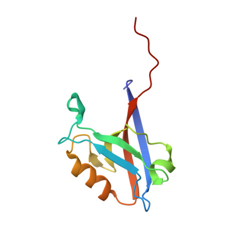
Interaction partners of PSD-93 studied by X-ray crystallography and fluorescence polarization spectroscopy.
Fiorentini, M., Bach, A., Stromgaard, K., Kastrup, J.S., Gajhede, M.(2013) Acta Crystallogr D Biol Crystallogr 69: 587-594
- PubMed: 23519667
- DOI: https://doi.org/10.1107/S0907444912051839
- Primary Citation of Related Structures:
4H11 - PubMed Abstract:
PSD-93 (chapsyn-110, DLG2) is a member of the family of membrane-associated guanylate kinase (MAGUK) proteins. The MAGUK proteins are involved in receptor localization and signalling pathways. The best characterized MAGUK protein, PSD-95, is known to be involved in NMDA receptor signalling via its PDZ domains. The PDZ domains of PSD-95 and PSD-93 are structurally very similar, but relatively little is known about the function of PSD-93. PSD-93 has been suggested to interact with GluD2 from the family of ionotropic glutamate receptors. Here, the interactions of four residues (GTSI) representing the extreme C-terminus of GluD2 with PSD-93 PDZ1 have been investigated in the crystalline phase. Two different binding modes of these residues were observed, suggesting that the peptide is not tightly bound to PSD-93 PDZ1. In accordance, the two N-terminal PSD-93 PDZ domains show no appreciable binding affinity for a GluD2-derived C-terminal octapeptide, whereas micromolar affinity was observed for a GluN2B-derived C-terminal octapeptide. This indicates that if present, the interactions between GluD2 and PSD-93 involve more than the extreme terminus of the receptor. In contrast, the tumour-suppressor protein SCRIB PDZ3 shows low micromolar affinity towards the GluD2-derived octapeptide, which is in agreement with previous findings using high-throughput assays.
Organizational Affiliation:
Department of Drug Design and Pharmacology, Faculty of Health and Medical Sciences, University of Copenhagen, Copenhagen, Denmark.