4GNG

Crystal Structure of NSD3 tandem PHD5-C5HCH domains complexed with H3K9me3 peptide


Experimental Data Snapshot

  • Method: X-RAY DIFFRACTION
  • Resolution: 1.73 Å
  • R-Value Free: 0.214 
  • R-Value Work: 0.181 
  • R-Value Observed: 0.183 

wwPDB Validation   3D Report Full Report


This is version 1.2 of the entry. See complete history


Literature

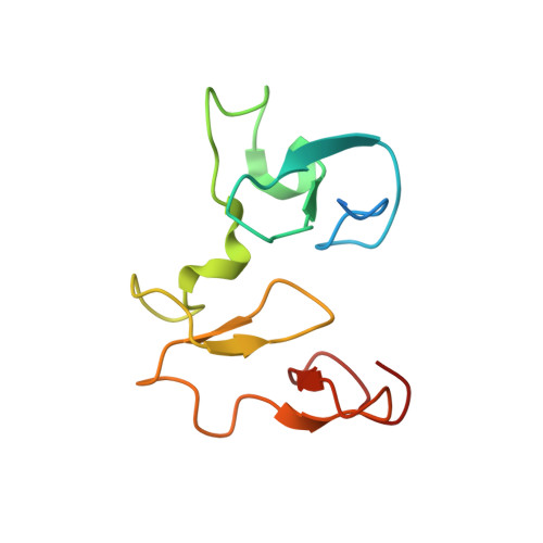
The methyltransferase NSD3 has chromatin-binding motifs, PHD5-C5HCH, that are distinct from other NSD (nuclear receptor SET domain) family members in their histone H3 recognition.

He, C.Li, F.Zhang, J.Wu, J.Shi, Y.

(2013) J Biol Chem 288: 4692-4703

  • DOI: https://doi.org/10.1074/jbc.M112.426148
  • Primary Citation of Related Structures:  
    4GND, 4GNE, 4GNF, 4GNG

  • PubMed Abstract: 

    The NSD (nuclear receptor SET domain-containing) family members, consisting of NSD1, NSD2 (MMSET/WHSC1), and NSD3 (WHSC1L1), are SET domain-containing methyltransferases and aberrant expression of each member has been implicated in multiple diseases. They have specific mono- and dimethylase activities for H3K36, whereas play nonredundant roles during development. Aside from the well characterized catalytic SET domain, NSD proteins have multiple potential chromatin-binding motifs that are clinically relevant, including the fifth plant homeodomain (PHD5) and the adjacent Cys-His-rich domain (C5HCH) located at the C terminus. Herein, we report the crystal structures of the PHD5-C5HCH module of NSD3, in the free state and in complex with H3(1-7) (H3 residues 1-7), H3(1-15) (H3 residues 1-15), and H3(1-15)K9me3 (H3 residues 1-15 with trimethylation on K9) peptides. These structures reveal that the PHD5 and C5HCH domains fold into a novel integrated PHD-PHD-like structural module with H3 peptide bound only on the surface of PHD5 and provide the molecular basis for the recognition of unmodified H3K4 and trimethylated H3K9 by NSD3 PHD5. Structural studies and binding assays show that differences exist in histone binding specificity of the PHD5 domain between three members of the NSD family. For NSD2, the PHD5-C5HCH:H3 N terminus interaction is largely conserved, although with a stronger preference for unmethylated H3K9 (H3K9me0) than trimethylated H3K9 (H3K9me3), and NSD1 PHD5-C5HCH does not bind to H3 peptides. Our results shed light on how NSD proteins that mediate H3K36 methylation are localized to specific genomic sites and provide implications for the mechanism of functional diversity of NSD proteins.


  • Organizational Affiliation

    Hefei National Laboratory for Physical Sciences at Microscale and School of Life Sciences, University of Science and Technology of China, Hefei, Anhui 230026, China.


Macromolecules
Find similar proteins by:  (by identity cutoff)  |  3D Structure
Entity ID: 1
MoleculeChains Sequence LengthOrganismDetailsImage
Histone-lysine N-methyltransferase NSD3A,
C [auth D]
107Homo sapiensMutation(s): 0 
Gene Names: WHSC1L1NSD3DC28
EC: 2.1.1.43
UniProt & NIH Common Fund Data Resources
Find proteins for Q9BZ95 (Homo sapiens)
Explore Q9BZ95 
Go to UniProtKB:  Q9BZ95
PHAROS:  Q9BZ95
GTEx:  ENSG00000147548 
Entity Groups  
Sequence Clusters30% Identity50% Identity70% Identity90% Identity95% Identity100% Identity
UniProt GroupQ9BZ95
Sequence Annotations
Expand
  • Reference Sequence

Find similar proteins by:  Sequence   |   3D Structure  

Entity ID: 2
MoleculeChains Sequence LengthOrganismDetailsImage
Histone H3.3B,
D [auth F]
15Homo sapiensMutation(s): 0 
UniProt & NIH Common Fund Data Resources
Find proteins for P84243 (Homo sapiens)
Explore P84243 
Go to UniProtKB:  P84243
PHAROS:  P84243
Entity Groups  
Sequence Clusters30% Identity50% Identity70% Identity90% Identity95% Identity100% Identity
UniProt GroupP84243
Sequence Annotations
Expand
  • Reference Sequence
Small Molecules
Ligands 2 Unique
IDChains Name / Formula / InChI Key2D Diagram3D Interactions
GOL
Query on GOL

Download Ideal Coordinates CCD File 
I [auth A],
J [auth B],
K [auth B]
GLYCEROL
C3 H8 O3
PEDCQBHIVMGVHV-UHFFFAOYSA-N
ZN
Query on ZN

Download Ideal Coordinates CCD File 
E [auth A]
F [auth A]
G [auth A]
H [auth A]
L [auth D]
E [auth A],
F [auth A],
G [auth A],
H [auth A],
L [auth D],
M [auth D],
N [auth D],
O [auth D]
ZINC ION
Zn
PTFCDOFLOPIGGS-UHFFFAOYSA-N
Modified Residues  1 Unique
IDChains TypeFormula2D DiagramParent
M3L
Query on M3L
B,
D [auth F]
L-PEPTIDE LINKINGC9 H21 N2 O2LYS
Experimental Data & Validation

Experimental Data

  • Method: X-RAY DIFFRACTION
  • Resolution: 1.73 Å
  • R-Value Free: 0.214 
  • R-Value Work: 0.181 
  • R-Value Observed: 0.183 
  • Space Group: P 21 21 21
Unit Cell:
Length ( Å )Angle ( ˚ )
a = 56.2α = 90
b = 59.96β = 90
c = 95.23γ = 90
Software Package:
Software NamePurpose
MOSFLMdata reduction
SCALAdata scaling
MOLREPphasing
REFMACrefinement
PDB_EXTRACTdata extraction
ADSCdata collection

Structure Validation

View Full Validation Report


Entry History 

Deposition Data

Revision History  (Full details and data files)

  • Version 1.0: 2013-01-02
    Type: Initial release
  • Version 1.1: 2013-09-04
    Changes: Database references
  • Version 1.2: 2023-11-08
    Changes: Data collection, Database references, Derived calculations, Refinement description