


4GF5
Crystal Structure of Calicheamicin Methyltransferase, CalS11
- PDB DOI: https://doi.org/10.2210/pdb4GF5/pdb
- Classification: TRANSFERASE
- Organism(s): Micromonospora echinospora
- Expression System: Escherichia coli
- Mutation(s): No
- Deposited: 2012-08-02 Released: 2012-08-22
Experimental Data Snapshot
- Method: X-RAY DIFFRACTION
- Resolution: 2.20 Å
- R-Value Free: 0.213
- R-Value Work: 0.159
- R-Value Observed: 0.159
Starting Model: experimental
View more details

This is version 1.2 of the entry. See complete history.
Literature
To be published.

Macromolecules
Find similar proteins by:
(by identity cutoff) | 3D Structure
Entity ID: 1 | |||||
|---|---|---|---|---|---|
| Molecule | Chains | Sequence Length | Organism | Details | Image |
| CalS11 | 257 | Micromonospora echinospora | Mutation(s): 0 Gene Names: AAM70337, calS11 | 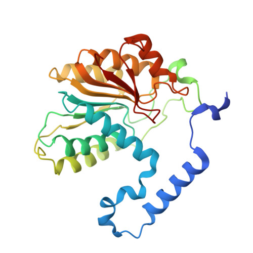 | |
UniProt | |||||
Find proteins for Q8KNF1 (Micromonospora echinospora) Explore Q8KNF1 Go to UniProtKB: Q8KNF1 | |||||
Entity Groups | |||||
| Sequence Clusters | 30% Identity50% Identity70% Identity90% Identity95% Identity100% Identity | ||||
| UniProt Group | Q8KNF1 | ||||
Sequence AnnotationsExpand | |||||
| |||||
Small Molecules
| Ligands 2 Unique | |||||
|---|---|---|---|---|---|
| ID | Chains | Name / Formula / InChI Key | 2D Diagram | 3D Interactions | |
| SAH Query on SAH | BA [auth F] BB [auth N] DB [auth O] EA [auth H] FB [auth P] | S-ADENOSYL-L-HOMOCYSTEINE C14 H20 N6 O5 S ZJUKTBDSGOFHSH-WFMPWKQPSA-N |  | ||
| SO4 Query on SO4 | AA [auth E] AB [auth M] CA [auth F] CB [auth N] DA [auth F] | SULFATE ION O4 S QAOWNCQODCNURD-UHFFFAOYSA-L |  | ||
Experimental Data & Validation
Experimental Data
- Method: X-RAY DIFFRACTION
- Resolution: 2.20 Å
- R-Value Free: 0.213
- R-Value Work: 0.159
- R-Value Observed: 0.159
- Space Group: P 1
Unit Cell:
| Length ( Å ) | Angle ( ˚ ) |
|---|---|
| a = 75.112 | α = 80.88 |
| b = 106.24 | β = 81.65 |
| c = 184.6 | γ = 70.02 |
| Software Name | Purpose |
|---|---|
| DENZO | data reduction |
| SCALEPACK | data scaling |
| PHASER | phasing |
| PHENIX | refinement |
| PDB_EXTRACT | data extraction |
| HKL-2000 | data collection |
| HKL-2000 | data reduction |
| HKL-2000 | data scaling |
| PHENIX | phasing |
Entry History
Deposition Data
- Released Date: 2012-08-22 Deposition Author(s): Helmich, K.E., Singh, S., Thorson, J.S., Phillips Jr., G.N., Enzyme Discovery for Natural Product Biosynthesis (NatPro), Center for Eukaryotic Structural Genomics (CESG)
Revision History (Full details and data files)
- Version 1.0: 2012-08-22
Type: Initial release - Version 1.1: 2017-11-15
Changes: Advisory, Refinement description - Version 1.2: 2023-09-13
Changes: Advisory, Data collection, Database references, Derived calculations, Refinement description
