4GDI

A subtype N10 neuraminidase-like protein of A/little yellow-shouldered bat/Guatemala/164/2009


Experimental Data Snapshot

  • Method: X-RAY DIFFRACTION
  • Resolution: 1.95 Å
  • R-Value Free: 0.223 
  • R-Value Work: 0.180 
  • R-Value Observed: 0.182 

wwPDB Validation   3D Report Full Report


Ligand Structure Quality Assessment 


This is version 2.1 of the entry. See complete history


Literature

Crystal structures of two subtype N10 neuraminidase-like proteins from bat influenza A viruses reveal a diverged putative active site.

Zhu, X.Yang, H.Guo, Z.Yu, W.Carney, P.J.Li, Y.Chen, L.M.Paulson, J.C.Donis, R.O.Tong, S.Stevens, J.Wilson, I.A.

(2012) Proc Natl Acad Sci U S A 109: 18903-18908

  • DOI: https://doi.org/10.1073/pnas.1212579109
  • Primary Citation of Related Structures:  
    4GDI, 4GDJ, 4GEZ

  • PubMed Abstract: 

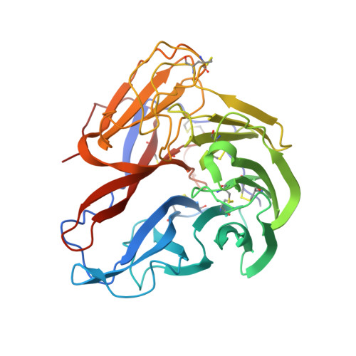
    Recently, we reported a unique influenza A virus subtype H17N10 from little yellow-shouldered bats. Its neuraminidase (NA) gene encodes a protein that appears to be highly divergent from all known influenza NAs and was assigned as a new subtype N10. To provide structural and functional insights on the bat H17N10 virus, X-ray structures were determined for N10 NA proteins from influenza A viruses A/little yellow-shouldered bat/Guatemala/164/2009 (GU09-164) in two crystal forms at 1.95 Å and 2.5 Å resolution and A/little yellow-shouldered bat/Guatemala/060/2010 (GU10-060) at 2.0 Å. The overall N10 structures are similar to each other and to other known influenza NA structures, with a single highly conserved calcium binding site in each monomer. However, the region corresponding to the highly conserved active site of influenza A N1-N9 NA subtypes and influenza B NA differs substantially. In particular, most of the amino acid residues required for NA activity are substituted, and the putative active site is much wider because of displacement of the 150-loop and 430-loop. These structural features and the fact that the recombinant N10 protein exhibits no, or extremely low, NA activity suggest that it may have a different function than the NA proteins of other influenza viruses. Accordingly, we propose that the N10 protein be termed an NA-like protein until its function is elucidated.


  • Organizational Affiliation

    Department of Molecular Biology, The Scripps Research Institute, La Jolla, CA 92037, USA.


Macromolecules
Find similar proteins by:  (by identity cutoff)  |  3D Structure
Entity ID: 1
MoleculeChains Sequence LengthOrganismDetailsImage
Neuraminidase
A, B, C, D, E
A, B, C, D, E, F
373Influenza A virus (A/little yellow-shouldered bat/Guatemala/164/2009(H17N10))Mutation(s): 0 
Gene Names: NA
UniProt
Find proteins for H6QM85 (Influenza A virus)
Explore H6QM85 
Go to UniProtKB:  H6QM85
Entity Groups  
Sequence Clusters30% Identity50% Identity70% Identity90% Identity95% Identity100% Identity
UniProt GroupH6QM85
Sequence Annotations
Expand
  • Reference Sequence
Oligosaccharides

Help

Entity ID: 2
MoleculeChains Length2D Diagram Glycosylation3D Interactions
beta-D-mannopyranose-(1-4)-2-acetamido-2-deoxy-beta-D-glucopyranose-(1-4)-[beta-L-fucopyranose-(1-3)][alpha-L-fucopyranose-(1-6)]2-acetamido-2-deoxy-beta-D-glucopyranose
G
5N-Glycosylation
Glycosylation Resources
GlyTouCan:  G31349EA
GlyCosmos:  G31349EA
Entity ID: 3
MoleculeChains Length2D Diagram Glycosylation3D Interactions
alpha-D-mannopyranose-(1-3)-beta-D-mannopyranose-(1-4)-2-acetamido-2-deoxy-beta-D-glucopyranose-(1-4)-[beta-L-fucopyranose-(1-3)][alpha-L-fucopyranose-(1-6)]2-acetamido-2-deoxy-beta-D-glucopyranose
H
6N-Glycosylation
Glycosylation Resources
GlyTouCan:  G88708XF
GlyCosmos:  G88708XF
GlyGen:  G88708XF
Entity ID: 4
MoleculeChains Length2D Diagram Glycosylation3D Interactions
beta-L-fucopyranose-(1-3)-[2-acetamido-2-deoxy-beta-D-glucopyranose-(1-4)][alpha-L-fucopyranose-(1-6)]2-acetamido-2-deoxy-beta-D-glucopyranose
I, K, M
4N-Glycosylation
Glycosylation Resources
GlyTouCan:  G62810GW
GlyCosmos:  G62810GW
Entity ID: 5
MoleculeChains Length2D Diagram Glycosylation3D Interactions
2-acetamido-2-deoxy-beta-D-glucopyranose-(1-4)-[alpha-L-fucopyranose-(1-6)]2-acetamido-2-deoxy-beta-D-glucopyranose
J
3N-Glycosylation
Glycosylation Resources
GlyTouCan:  G21290RB
GlyCosmos:  G21290RB
GlyGen:  G21290RB
Entity ID: 6
MoleculeChains Length2D Diagram Glycosylation3D Interactions
alpha-L-fucopyranose-(1-6)-2-acetamido-2-deoxy-beta-D-glucopyranose
L
2N-Glycosylation
Glycosylation Resources
GlyTouCan:  G86851RC
GlyCosmos:  G86851RC
GlyGen:  G86851RC
Entity ID: 7
MoleculeChains Length2D Diagram Glycosylation3D Interactions
beta-L-fucopyranose-(1-3)-[2-acetamido-2-deoxy-beta-D-glucopyranose-(1-4)]2-acetamido-2-deoxy-beta-D-glucopyranose
N
3N-Glycosylation
Glycosylation Resources
GlyTouCan:  G46874CY
GlyCosmos:  G46874CY
GlyGen:  G46874CY
Small Molecules
Ligands 4 Unique
IDChains Name / Formula / InChI Key2D Diagram3D Interactions
NAG
Query on NAG

Download Ideal Coordinates CCD File 
DB [auth F]
EB [auth F]
IA [auth C]
JA [auth C]
O [auth A]
DB [auth F],
EB [auth F],
IA [auth C],
JA [auth C],
O [auth A],
P [auth A],
PA [auth D],
Q [auth A],
QA [auth D],
WA [auth E],
X [auth B],
XA [auth E],
Y [auth B]
2-acetamido-2-deoxy-beta-D-glucopyranose
C8 H15 N O6
OVRNDRQMDRJTHS-FMDGEEDCSA-N
GOL
Query on GOL

Download Ideal Coordinates CCD File 
AB [auth E]
BA [auth B]
BB [auth E]
CA [auth B]
CB [auth E]
AB [auth E],
BA [auth B],
BB [auth E],
CA [auth B],
CB [auth E],
HB [auth F],
IB [auth F],
JB [auth F],
KB [auth F],
MA [auth C],
S [auth A],
T [auth A],
TA [auth D],
U [auth A]
GLYCEROL
C3 H8 O3
PEDCQBHIVMGVHV-UHFFFAOYSA-N
NO3
Query on NO3

Download Ideal Coordinates CCD File 
DA [auth B]
EA [auth B]
FA [auth B]
GA [auth B]
HA [auth B]
DA [auth B],
EA [auth B],
FA [auth B],
GA [auth B],
HA [auth B],
LB [auth F],
MB [auth F],
NA [auth C],
NB [auth F],
OA [auth C],
UA [auth D],
V [auth A],
VA [auth D],
W [auth A]
NITRATE ION
N O3
NHNBFGGVMKEFGY-UHFFFAOYSA-N
CA
Query on CA

Download Ideal Coordinates CCD File 
AA [auth B]
FB [auth F]
GB [auth F]
KA [auth C]
LA [auth C]
AA [auth B],
FB [auth F],
GB [auth F],
KA [auth C],
LA [auth C],
R [auth A],
RA [auth D],
SA [auth D],
YA [auth E],
Z [auth B],
ZA [auth E]
CALCIUM ION
Ca
BHPQYMZQTOCNFJ-UHFFFAOYSA-N
Experimental Data & Validation

Experimental Data

  • Method: X-RAY DIFFRACTION
  • Resolution: 1.95 Å
  • R-Value Free: 0.223 
  • R-Value Work: 0.180 
  • R-Value Observed: 0.182 
  • Space Group: I 4
Unit Cell:
Length ( Å )Angle ( ˚ )
a = 176.324α = 90
b = 176.324β = 90
c = 193.313γ = 90
Software Package:
Software NamePurpose
PHENIXrefinement

Structure Validation

View Full Validation Report


Ligand Structure Quality Assessment 


Entry History 

Deposition Data

Revision History  (Full details and data files)

  • Version 1.0: 2012-09-26
    Type: Initial release
  • Version 1.1: 2012-10-10
    Changes: Database references
  • Version 1.2: 2012-11-28
    Changes: Database references
  • Version 1.3: 2014-12-17
    Changes: Data collection
  • Version 2.0: 2020-07-29
    Type: Remediation
    Reason: Carbohydrate remediation
    Changes: Atomic model, Data collection, Database references, Derived calculations, Structure summary
  • Version 2.1: 2023-09-13
    Changes: Data collection, Database references, Refinement description, Structure summary