4GCZ

Structure of a blue-light photoreceptor


Experimental Data Snapshot

  • Method: X-RAY DIFFRACTION
  • Resolution: 2.30 Å
  • R-Value Free: 0.217 
  • R-Value Work: 0.179 
  • R-Value Observed: 0.181 

wwPDB Validation   3D Report Full Report


Ligand Structure Quality Assessment 


This is version 1.3 of the entry. See complete history


Literature

Full-length structure of a sensor histidine kinase pinpoints coaxial coiled coils as signal transducers and modulators.

Diensthuber, R.P.Bommer, M.Gleichmann, T.Moglich, A.

(2013) Structure 21: 1127-1136

  • DOI: https://doi.org/10.1016/j.str.2013.04.024
  • Primary Citation of Related Structures:  
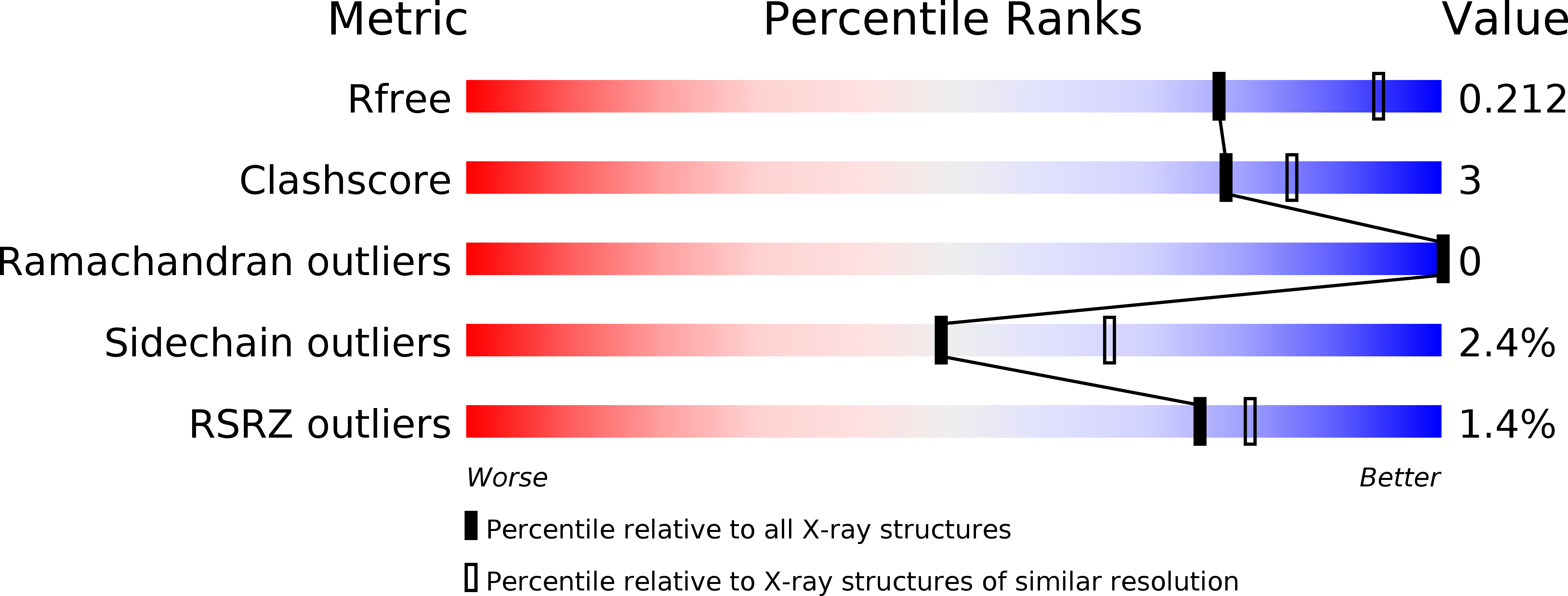
    4GCZ

  • PubMed Abstract: 

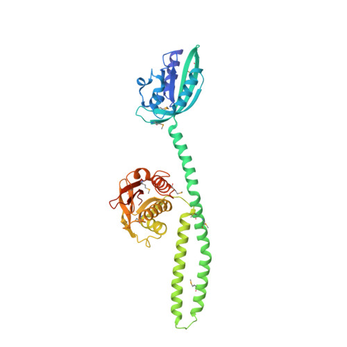
    Two-component systems (TCSs), which comprise sensor histidine kinases (SHK) and response-regulator proteins, represent the predominant strategy by which prokaryotes sense and respond to a changing environment. Despite paramount biological importance, a dearth exists of intact SHK structures containing both sensor and effector modules. Here, we report the full-length crystal structure of the engineered, dimeric, blue-light-regulated SHK YF1 at 2.3 Å resolution, in which two N-terminal light-oxygen-voltage (LOV) photosensors are connected by a coiled coil to the C-terminal effector modules. A second coaxial coiled coil derived from the N-termini of the LOV photosensors and inserted between them crucially modulates light regulation: single mutations within this coiled coil attenuate or even invert the signal response of the TCS. Structural motifs identified in YF1 recur in signal receptors, and the underlying signaling principles and mechanisms may be widely shared between soluble and transmembrane, prokaryotic, and eukaryotic signal receptors of diverse biological activity.


  • Organizational Affiliation

    Biophysikalische Chemie, Institut für Biologie, Humboldt-Universität zu Berlin, 10115 Berlin, Germany. ralph.diensthuber@hu-berlin.de


Macromolecules
Find similar proteins by:  (by identity cutoff)  |  3D Structure
Entity ID: 1
MoleculeChains Sequence LengthOrganismDetailsImage
Blue-light photoreceptor, Sensor protein fixL
A, B
385Bacillus subtilis subsp. subtilis str. 168Bradyrhizobium diazoefficiens USDA 110
This entity is chimeric
Mutation(s): 0 
Gene Names: pfyPytvABSU30340fixLbll2760
UniProt
Find proteins for P23222 (Bradyrhizobium diazoefficiens (strain JCM 10833 / BCRC 13528 / IAM 13628 / NBRC 14792 / USDA 110))
Explore P23222 
Go to UniProtKB:  P23222
Find proteins for O34627 (Bacillus subtilis (strain 168))
Explore O34627 
Go to UniProtKB:  O34627
Entity Groups  
Sequence Clusters30% Identity50% Identity70% Identity90% Identity95% Identity100% Identity
UniProt GroupsP23222O34627
Sequence Annotations
Expand
  • Reference Sequence
Small Molecules
Ligands 3 Unique
IDChains Name / Formula / InChI Key2D Diagram3D Interactions
FMN
Query on FMN

Download Ideal Coordinates CCD File 
C [auth A],
I [auth B]
FLAVIN MONONUCLEOTIDE
C17 H21 N4 O9 P
FVTCRASFADXXNN-SCRDCRAPSA-N
ADP
Query on ADP

Download Ideal Coordinates CCD File 
D [auth A]ADENOSINE-5'-DIPHOSPHATE
C10 H15 N5 O10 P2
XTWYTFMLZFPYCI-KQYNXXCUSA-N
SO4
Query on SO4

Download Ideal Coordinates CCD File 
E [auth A]
F [auth A]
G [auth A]
H [auth A]
J [auth B]
E [auth A],
F [auth A],
G [auth A],
H [auth A],
J [auth B],
K [auth B],
L [auth B],
M [auth B],
N [auth B],
O [auth B]
SULFATE ION
O4 S
QAOWNCQODCNURD-UHFFFAOYSA-L
Modified Residues  1 Unique
IDChains TypeFormula2D DiagramParent
MSE
Query on MSE
A, B
L-PEPTIDE LINKINGC5 H11 N O2 SeMET
Experimental Data & Validation

Experimental Data

  • Method: X-RAY DIFFRACTION
  • Resolution: 2.30 Å
  • R-Value Free: 0.217 
  • R-Value Work: 0.179 
  • R-Value Observed: 0.181 
  • Space Group: P 65 2 2
Unit Cell:
Length ( Å )Angle ( ˚ )
a = 105.216α = 90
b = 105.216β = 90
c = 441.958γ = 120
Software Package:
Software NamePurpose
XSCALEdata scaling
PHENIXrefinement
PDB_EXTRACTdata extraction
MxCuBEdata collection
XDSdata reduction
XDSdata scaling
PHASERphasing

Structure Validation

View Full Validation Report


Ligand Structure Quality Assessment 


Entry History 

Deposition Data

Revision History  (Full details and data files)

  • Version 1.0: 2013-06-19
    Type: Initial release
  • Version 1.1: 2013-07-10
    Changes: Database references
  • Version 1.2: 2013-07-24
    Changes: Database references
  • Version 1.3: 2017-08-23
    Changes: Advisory, Refinement description, Source and taxonomy