4G6T

Structure of the HopA1-SchA Chaperone-Effector Complex


Experimental Data Snapshot

  • Method: X-RAY DIFFRACTION
  • Resolution: 1.56 Å
  • R-Value Free: 0.230 
  • R-Value Work: 0.209 
  • R-Value Observed: 0.211 

wwPDB Validation   3D Report Full Report


This is version 1.1 of the entry. See complete history


Literature

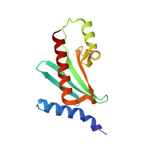
Structure of the HopA1(21-102)-ShcA chaperone-effector complex of Pseudomonas syringae reveals conservation of a virulence factor binding motif from animal to plant pathogens.

Janjusevic, R.Quezada, C.M.Small, J.Stebbins, C.E.

(2013) J Bacteriol 195: 658-664

  • DOI: https://doi.org/10.1128/JB.01621-12
  • Primary Citation of Related Structures:  
    4G6T

  • PubMed Abstract: 

    Pseudomonas syringae injects numerous bacterial proteins into host plant cells through a type 3 secretion system (T3SS). One of the first such bacterial effectors discovered, HopA1, is a protein that has unknown functions in the host cell but possesses close homologs that trigger the plant hypersensitive response in resistant strains. Like the virulence factors in many bacterial pathogens of animals, HopA1 depends upon a cognate chaperone in order to be effectively translocated by the P. syringae T3SS. Herein, we report the crystal structure of a complex of HopA1(21-102) with its chaperone, ShcA, determined to 1.56-Å resolution. The structure reveals that three key features of the chaperone-effector interactions found in animal pathogens are preserved in the Gram-negative pathogens of plants, namely, (i) the interaction of the chaperone with a nonglobular polypeptide of the effector, (ii) an interaction centered on the so-called β-motif, and (iii) the presence of a conserved hydrophobic patch in the chaperone that recognizes the β-motif. Structure-based mutagenesis and biochemical studies have established that the β-motif is critical for the stability of this complex. Overall, these results show that the β-motif interactions are broadly conserved in bacterial pathogens utilizing T3SSs, spanning an interkingdom host range.


  • Organizational Affiliation

    Laboratory of Structural Microbiology, The Rockefeller University, New York, NY, USA.


Macromolecules
Find similar proteins by:  (by identity cutoff)  |  3D Structure
Entity ID: 1
MoleculeChains Sequence LengthOrganismDetailsImage
Type III chaperone protein ShcA128Pseudomonas syringae pv. tomato str. DC3000Mutation(s): 0 
Gene Names: shcAPSPTO_5353
UniProt
Find proteins for Q87UE6 (Pseudomonas syringae pv. tomato (strain ATCC BAA-871 / DC3000))
Explore Q87UE6 
Go to UniProtKB:  Q87UE6
Entity Groups  
Sequence Clusters30% Identity50% Identity70% Identity90% Identity95% Identity100% Identity
UniProt GroupQ87UE6
Sequence Annotations
Expand
  • Reference Sequence
Find similar proteins by:  (by identity cutoff)  |  3D Structure
Entity ID: 2
MoleculeChains Sequence LengthOrganismDetailsImage
Type III effector HopA182Pseudomonas syringae pv. tomato str. DC3000Mutation(s): 0 
Gene Names: hopA1PSPTO_5354
UniProt
Find proteins for Q87UE5 (Pseudomonas syringae pv. tomato (strain ATCC BAA-871 / DC3000))
Explore Q87UE5 
Go to UniProtKB:  Q87UE5
Entity Groups  
Sequence Clusters30% Identity50% Identity70% Identity90% Identity95% Identity100% Identity
UniProt GroupQ87UE5
Sequence Annotations
Expand
  • Reference Sequence
Experimental Data & Validation

Experimental Data

  • Method: X-RAY DIFFRACTION
  • Resolution: 1.56 Å
  • R-Value Free: 0.230 
  • R-Value Work: 0.209 
  • R-Value Observed: 0.211 
  • Space Group: P 65 2 2
Unit Cell:
Length ( Å )Angle ( ˚ )
a = 67.832α = 90
b = 67.832β = 90
c = 182.887γ = 120
Software Package:
Software NamePurpose
REFMACrefinement
HKL-2000data reduction
HKL-2000data scaling

Structure Validation

View Full Validation Report


Entry History 

Deposition Data

Revision History  (Full details and data files)

  • Version 1.0: 2013-06-05
    Type: Initial release
  • Version 1.1: 2024-02-28
    Changes: Data collection, Database references