4G1Q

Crystal structure of HIV-1 reverse transcriptase (RT) in complex with Rilpivirine (TMC278, Edurant), a non-nucleoside rt-inhibiting drug


Experimental Data Snapshot

  • Method: X-RAY DIFFRACTION
  • Resolution: 1.51 Å
  • R-Value Free: 0.193 
  • R-Value Work: 0.154 
  • R-Value Observed: 0.155 

wwPDB Validation   3D Report Full Report


This is version 1.4 of the entry. See complete history


Literature

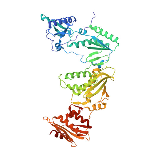
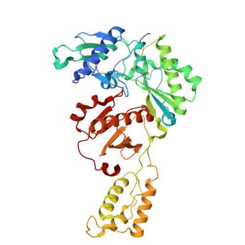
Snapshot of the equilibrium dynamics of a drug bound to HIV-1 reverse transcriptase.

Kuroda, D.G.Bauman, J.D.Challa, J.R.Patel, D.Troxler, T.Das, K.Arnold, E.Hochstrasser, R.M.

(2013) Nat Chem 5: 174-181

  • DOI: https://doi.org/10.1038/nchem.1559
  • Primary Citation of Related Structures:  
    4G1Q

  • PubMed Abstract: 

    The anti-AIDS drug rilpivirine undergoes conformational changes to bind HIV-1 reverse transcriptase (RT), which is an essential enzyme for the replication of HIV. These changes allow it to retain potency against mutations that otherwise would render the enzyme resistant. Here we report that water molecules play an essential role in this binding process. Femtosecond experiments and theory expose the molecular level dynamics of rilpivirine bound to HIV-1 RT. Two nitrile substituents, one on each arm of the drug, are used as vibrational probes of the structural dynamics within the binding pocket. Two-dimensional vibrational echo spectroscopy reveals that one nitrile group is unexpectedly hydrogen-bonded to a mobile water molecule, not identified in previous X-ray structures. Ultrafast nitrile-water dynamics are confirmed by simulations. A higher (1.51 Å) resolution X-ray structure also reveals a water-drug interaction network. Maintenance of a crucial anchoring hydrogen bond may help retain the potency of rilpivirine against pocket mutations despite the structural variations they cause.


  • Organizational Affiliation

    Ultrafast Optical Processes Laboratory, Department of Chemistry, University of Pennsylvania, Pennsylvania 19067, USA.


Macromolecules
Find similar proteins by:  (by identity cutoff)  |  3D Structure
Entity ID: 1
MoleculeChains Sequence LengthOrganismDetailsImage
Reverse transcriptase/ribonuclease H557Human immunodeficiency virus type 1 BH10Mutation(s): 3 
Gene Names: gag-pol
EC: 2.7.7.49 (PDB Primary Data), 2.7.7.7 (PDB Primary Data), 3.1.26.13 (PDB Primary Data)
UniProt
Find proteins for P03366 (Human immunodeficiency virus type 1 group M subtype B (isolate BH10))
Explore P03366 
Go to UniProtKB:  P03366
Entity Groups  
Sequence Clusters30% Identity50% Identity70% Identity90% Identity95% Identity100% Identity
UniProt GroupP03366
Sequence Annotations
Expand
  • Reference Sequence
Find similar proteins by:  (by identity cutoff)  |  3D Structure
Entity ID: 2
MoleculeChains Sequence LengthOrganismDetailsImage
p51 RT428Human immunodeficiency virus type 1 BH10Mutation(s): 1 
Gene Names: gag-pol
EC: 2.7.7.49 (PDB Primary Data), 2.7.7.7 (PDB Primary Data), 3.1.26.13 (PDB Primary Data)
UniProt
Find proteins for P03366 (Human immunodeficiency virus type 1 group M subtype B (isolate BH10))
Explore P03366 
Go to UniProtKB:  P03366
Entity Groups  
Sequence Clusters30% Identity50% Identity70% Identity90% Identity95% Identity100% Identity
UniProt GroupP03366
Sequence Annotations
Expand
  • Reference Sequence
Small Molecules
Ligands 4 Unique
IDChains Name / Formula / InChI Key2D Diagram3D Interactions
T27
Query on T27

Download Ideal Coordinates CCD File 
C [auth A]4-{[4-({4-[(E)-2-cyanoethenyl]-2,6-dimethylphenyl}amino)pyrimidin-2-yl]amino}benzonitrile
C22 H18 N6
YIBOMRUWOWDFLG-ONEGZZNKSA-N
SO4
Query on SO4

Download Ideal Coordinates CCD File 
D [auth A]
E [auth A]
F [auth A]
G [auth A]
K [auth B]
D [auth A],
E [auth A],
F [auth A],
G [auth A],
K [auth B],
L [auth B]
SULFATE ION
O4 S
QAOWNCQODCNURD-UHFFFAOYSA-L
EDO
Query on EDO

Download Ideal Coordinates CCD File 
H [auth A],
I [auth A],
M [auth B],
N [auth B],
O [auth B]
1,2-ETHANEDIOL
C2 H6 O2
LYCAIKOWRPUZTN-UHFFFAOYSA-N
MG
Query on MG

Download Ideal Coordinates CCD File 
J [auth A]MAGNESIUM ION
Mg
JLVVSXFLKOJNIY-UHFFFAOYSA-N
Binding Affinity Annotations 
IDSourceBinding Affinity
T27 BindingDB:  4G1Q IC50: min: 38, max: 1.00e+4 (nM) from 3 assay(s)
Experimental Data & Validation

Experimental Data

  • Method: X-RAY DIFFRACTION
  • Resolution: 1.51 Å
  • R-Value Free: 0.193 
  • R-Value Work: 0.154 
  • R-Value Observed: 0.155 
  • Space Group: C 1 2 1
Unit Cell:
Length ( Å )Angle ( ˚ )
a = 162.713α = 90
b = 72.517β = 100.84
c = 109.514γ = 90
Software Package:
Software NamePurpose
ADSCdata collection
PHENIXmodel building
PHENIXrefinement
HKL-2000data reduction
HKL-2000data scaling
PHENIXphasing

Structure Validation

View Full Validation Report


Entry History 

Deposition Data

Revision History  (Full details and data files)

  • Version 1.0: 2013-02-06
    Type: Initial release
  • Version 1.1: 2013-03-06
    Changes: Database references
  • Version 1.2: 2015-06-17
    Changes: Non-polymer description
  • Version 1.3: 2018-01-24
    Changes: Structure summary
  • Version 1.4: 2023-09-13
    Changes: Data collection, Database references, Derived calculations, Refinement description