


Crystal structure of HmoB with Heme
Park, S.To be published.
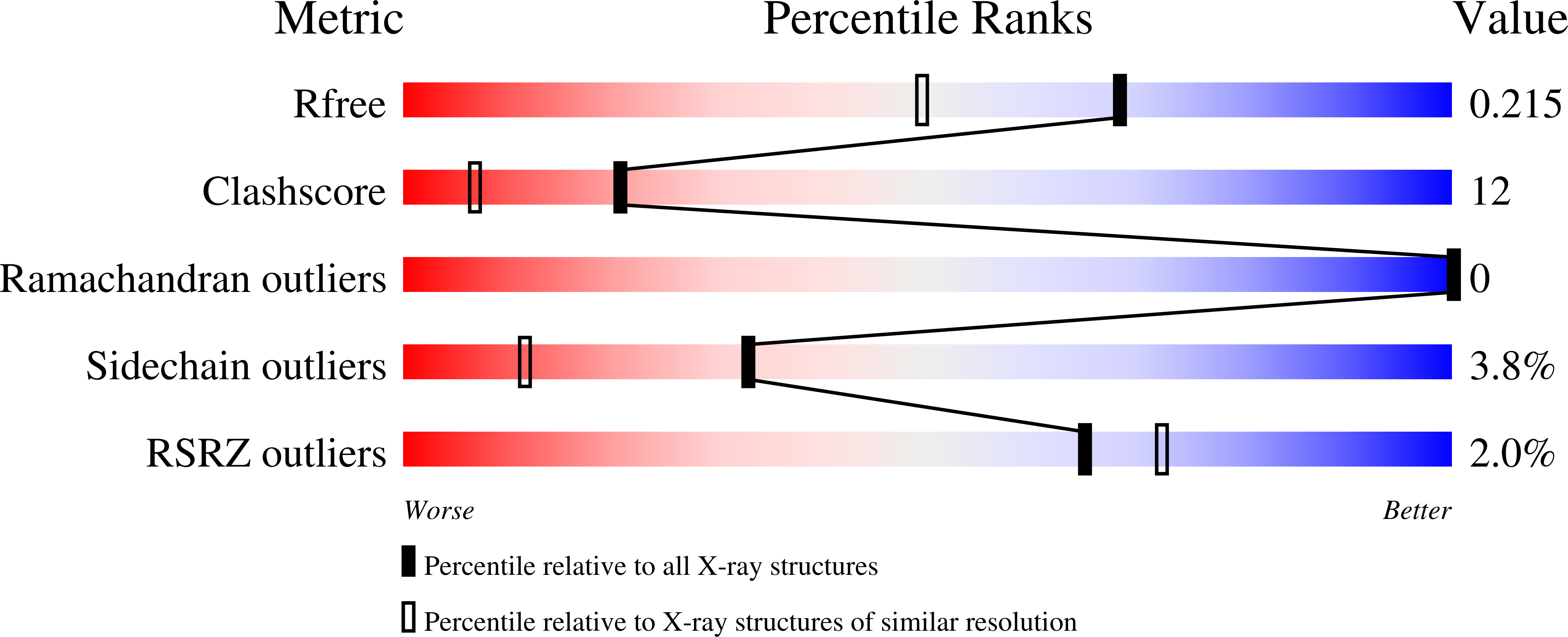
Experimental Data Snapshot
Entity ID: 1 | |||||
|---|---|---|---|---|---|
| Molecule | Chains | Sequence Length | Organism | Details | Image |
| Putative uncharacterized protein yhgC | 166 | Bacillus spizizenii str. W23 | Mutation(s): 0 Gene Names: yhgC, BSUW23_05110 |  | |
UniProt | |||||
Find proteins for E0TXX3 (Bacillus spizizenii (strain ATCC 23059 / NRRL B-14472 / W23)) Explore E0TXX3 Go to UniProtKB: E0TXX3 | |||||
Entity Groups | |||||
| Sequence Clusters | 30% Identity50% Identity70% Identity90% Identity95% Identity100% Identity | ||||
| UniProt Group | E0TXX3 | ||||
Sequence AnnotationsExpand | |||||
| |||||
| Ligands 1 Unique | |||||
|---|---|---|---|---|---|
| ID | Chains | Name / Formula / InChI Key | 2D Diagram | 3D Interactions | |
| HEM Query on HEM | G [auth A] H [auth B] I [auth C] J [auth D] K [auth E] | PROTOPORPHYRIN IX CONTAINING FE C34 H32 Fe N4 O4 KABFMIBPWCXCRK-RGGAHWMASA-L |  | ||
| Length ( Å ) | Angle ( ˚ ) |
|---|---|
| a = 70.821 | α = 90 |
| b = 117.519 | β = 91.22 |
| c = 70.847 | γ = 90 |
| Software Name | Purpose |
|---|---|
| HKL-2000 | data collection |
| CCP4 | model building |
| REFMAC | refinement |
| HKL-2000 | data reduction |
| HKL-2000 | data scaling |
| CCP4 | phasing |
RCSB PDB (citation) is hosted by
RCSB PDB is a member of the

