4FEU

Crystal structure of the aminoglycoside phosphotransferase APH(3')-Ia, with substrate kanamycin and small molecule inhibitor anthrapyrazolone SP600125


Experimental Data Snapshot

  • Method: X-RAY DIFFRACTION
  • Resolution: 2.37 Å
  • R-Value Free: 0.215 
  • R-Value Work: 0.160 
  • R-Value Observed: 0.163 

wwPDB Validation   3D Report Full Report


Ligand Structure Quality Assessment 


This is version 1.6 of the entry. See complete history


Literature

Structure-guided optimization of protein kinase inhibitors reverses aminoglycoside antibiotic resistance.

Stogios, P.J.Spanogiannopoulos, P.Evdokimova, E.Egorova, O.Shakya, T.Todorovic, N.Capretta, A.Wright, G.D.Savchenko, A.

(2013) Biochem J 454: 191-200

  • DOI: https://doi.org/10.1042/BJ20130317
  • Primary Citation of Related Structures:  
    4EJ7, 4FEU, 4FEV, 4FEW, 4FEX, 4GKH, 4GKI

  • PubMed Abstract: 

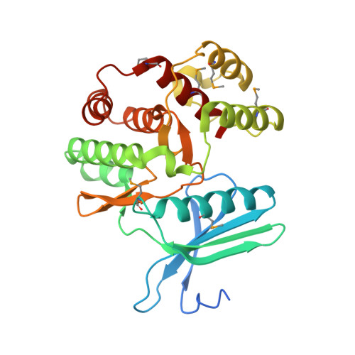
    Activity of the aminoglycoside phosphotransferase APH(3')-Ia leads to resistance to aminoglycoside antibiotics in pathogenic Gram-negative bacteria, and contributes to the clinical obsolescence of this class of antibiotics. One strategy to rescue compromised antibiotics such as aminoglycosides is targeting the enzymes that confer resistance with small molecules. We demonstrated previously that ePK (eukaryotic protein kinase) inhibitors could inhibit APH enzymes, owing to the structural similarity between these two enzyme families. However, limited structural information of enzyme-inhibitor complexes hindered interpretation of the results. In addition, cross-reactivity of compounds between APHs and ePKs represents an obstacle to their use as aminoglycoside adjuvants to rescue aminoglycoside antibiotic activity. In the present study, we structurally and functionally characterize inhibition of APH(3')-Ia by three diverse chemical scaffolds, anthrapyrazolone, 4-anilinoquinazoline and PP (pyrazolopyrimidine), and reveal distinctions in the binding mode of anthrapyrazolone and PP compounds to APH(3')-Ia compared with ePKs. Using this observation, we identify PP derivatives that select against ePKs, attenuate APH(3')-Ia activity and rescue aminoglycoside antibiotic activity against a resistant Escherichia coli strain. The structures described in the present paper and the inhibition studies provide an important opportunity for structure-based design of compounds to target aminoglycoside phosphotransferases for inhibition, potentially overcoming this form of antibiotic resistance.


  • Organizational Affiliation

    Department of Chemical Engineering and Applied Chemistry, University of Toronto, Ontario, Canada.


Macromolecules
Find similar proteins by:  (by identity cutoff)  |  3D Structure
Entity ID: 1
MoleculeChains Sequence LengthOrganismDetailsImage
Aminoglycoside 3'-phosphotransferase AphA1-IAB
A, B, C, D, E
A, B, C, D, E, F
272Acinetobacter baumannii AYEMutation(s): 0 
Gene Names: ABAYE3578APHA1-IAB
EC: 2.7.1.95
Entity Groups  
Sequence Clusters30% Identity50% Identity70% Identity90% Identity95% Identity100% Identity
Sequence Annotations
Expand
  • Reference Sequence
Small Molecules
Ligands 3 Unique
IDChains Name / Formula / InChI Key2D Diagram3D Interactions
KAN
Query on KAN

Download Ideal Coordinates CCD File 
G [auth A]
J [auth B]
L [auth C]
P [auth D]
R [auth E]
G [auth A],
J [auth B],
L [auth C],
P [auth D],
R [auth E],
T [auth F]
KANAMYCIN A
C18 H36 N4 O11
SBUJHOSQTJFQJX-NOAMYHISSA-N
537
Query on 537

Download Ideal Coordinates CCD File 
H [auth A],
M [auth C],
S [auth E]
2,6-DIHYDROANTHRA/1,9-CD/PYRAZOL-6-ONE
C14 H8 N2 O
ACPOUJIDANTYHO-UHFFFAOYSA-N
ACT
Query on ACT

Download Ideal Coordinates CCD File 
I [auth A]
K [auth B]
N [auth C]
O [auth C]
Q [auth D]
I [auth A],
K [auth B],
N [auth C],
O [auth C],
Q [auth D],
U [auth F]
ACETATE ION
C2 H3 O2
QTBSBXVTEAMEQO-UHFFFAOYSA-M
Modified Residues  1 Unique
IDChains TypeFormula2D DiagramParent
MSE
Query on MSE
A, B, C, D, E
A, B, C, D, E, F
L-PEPTIDE LINKINGC5 H11 N O2 SeMET
Experimental Data & Validation

Experimental Data

  • Method: X-RAY DIFFRACTION
  • Resolution: 2.37 Å
  • R-Value Free: 0.215 
  • R-Value Work: 0.160 
  • R-Value Observed: 0.163 
  • Space Group: P 1
  • Diffraction Data: https://doi.org/10.18430/M34FEU
Unit Cell:
Length ( Å )Angle ( ˚ )
a = 57.752α = 61.21
b = 94.161β = 73.11
c = 96.766γ = 87.36
Software Package:
Software NamePurpose
HKL-3000data collection
PHENIXmodel building
BUSTERrefinement
HKL-2000data reduction
HKL-2000data scaling
PHENIXphasing

Structure Validation

View Full Validation Report


Ligand Structure Quality Assessment 


Entry History 

Revision History  (Full details and data files)

  • Version 1.0: 2012-06-20
    Type: Initial release
  • Version 1.1: 2013-03-20
    Changes: Other
  • Version 1.2: 2013-06-26
    Changes: Database references
  • Version 1.3: 2013-08-07
    Changes: Database references
  • Version 1.4: 2013-09-04
    Changes: Database references
  • Version 1.5: 2023-09-13
    Changes: Data collection, Database references, Derived calculations, Refinement description
  • Version 1.6: 2023-12-06
    Changes: Data collection