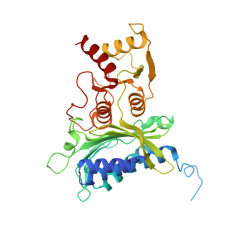
Conformational transition of fructose-1,6-bisphosphatase: structure comparison between the AMP complex (T form) and the fructose 6-phosphate complex (R form).
Ke, H.M., Liang, J.Y., Zhang, Y.P., Lipscomb, W.N.(1991) Biochemistry 30: 4412-4420
- PubMed: 1850623
- DOI: https://doi.org/10.1021/bi00232a007
- Primary Citation of Related Structures:
4FBP - PubMed Abstract:
A structure of the neutral form of fructose-1,6-bisphosphatase complexed with AMP has been determined by the molecular replacement method and refined at a 2.5-A resolution to a crystallographic R factor of 0.169. The root-mean-square errors of the structure from standard geometry are 0.013 A for bond lengths and 2.99 degrees for bond angles. Comparison of the AMP complex with the F6P complex shows that dimer C3-C4 twists about 19 degrees about a molecular 2-fold axis when dimers C1-C2 of the R and T forms of the enzyme are superimposed one another and that a slight shift of about 1 A of the AMP domain partially compensates this twist. The R to T transition of the enzyme does not significantly change the conformation of the F6P-binding site. However, residues at the divalent metal site and the AMP site show significant positional shifts. If these results can be extended to substrate in place of F6P, they suggest that regulation of the enzyme by AMP may occur partly through effects on metal-ion affinity or position. AMP binds to the same sites of the T and R forms, but only half-occupancy was observed in the alkaline R form. Sequential binding of AMP, at least in pairs, is suggested as the unligated R form is converted to the T form. Two possible pathways are suggested for allosteric communication over about 28 A between the AMP site and the active site: one via helices H1, H2, and H3 and another via the eight-stranded beta-sheet.(ABSTRACT TRUNCATED AT 250 WORDS)
Organizational Affiliation:
Gibbs Chemical Laboratory, Harvard University, Cambridge, Massachusetts 02138.