


Crystal structure of H74E synechocystis sp. pcya sp. PCYA
Kabasakal, B.V., Gae, D.D., Fisher, A.J.To be published.
Experimental Data Snapshot
Entity ID: 1 | |||||
|---|---|---|---|---|---|
| Molecule | Chains | Sequence Length | Organism | Details | Image |
| Phycocyanobilin:ferredoxin oxidoreductase | 248 | Synechocystis sp. PCC 6803 substr. Kazusa | Mutation(s): 1 Gene Names: pcyA, slr0116 EC: 1.3.7.5 | 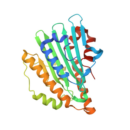 | |
UniProt | |||||
Find proteins for Q55891 (Synechocystis sp. (strain PCC 6803 / Kazusa)) Explore Q55891 Go to UniProtKB: Q55891 | |||||
Entity Groups | |||||
| Sequence Clusters | 30% Identity50% Identity70% Identity90% Identity95% Identity100% Identity | ||||
| UniProt Group | Q55891 | ||||
Sequence AnnotationsExpand | |||||
| |||||
| Ligands 2 Unique | |||||
|---|---|---|---|---|---|
| ID | Chains | Name / Formula / InChI Key | 2D Diagram | 3D Interactions | |
| BLA Query on BLA | B [auth A] | BILIVERDINE IX ALPHA C33 H34 N4 O6 GWZYPXHJIZCRAJ-SRVCBVSDSA-N |  | ||
| CL Query on CL | C [auth A], D [auth A] | CHLORIDE ION Cl VEXZGXHMUGYJMC-UHFFFAOYSA-M |  | ||
| Length ( Å ) | Angle ( ˚ ) |
|---|---|
| a = 70.638 | α = 90 |
| b = 95.53 | β = 90 |
| c = 42.767 | γ = 90 |
| Software Name | Purpose |
|---|---|
| Blu-Ice | data collection |
| PHASER | phasing |
| REFMAC | refinement |
| XDS | data reduction |
| XSCALE | data scaling |
RCSB PDB (citation) is hosted by
RCSB PDB is a member of the

