Molecular determinants of inactivation of the resuscitation promoting factor B from Mycobacterium tuberculosis.
Ruggiero, A., Marchant, J., Squeglia, F., Makarov, V., De Simone, A., Berisio, R.(2013) J Biomol Struct Dyn 31: 195-205
- PubMed: 22831279
- DOI: https://doi.org/10.1080/07391102.2012.698243
- Primary Citation of Related Structures:
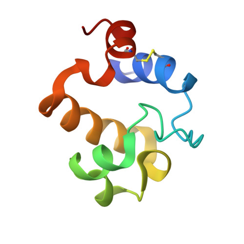
4EMN - PubMed Abstract:
Inactivation of revival of Mycobacterium tuberculosis from dormancy is one of the main goals of the WHO Global Plan to stop tuberculosis (TB) 2011-2015, given the huge reservoir of latently infected individuals. This process requires a group of secreted proteins, denoted as resuscitation-promoting factors (Rpfs). Of these, RpfB is the sole member indispensable for resuscitation in vivo. The first class of inhibitors of RpfB was identified among 2-nitrophenylthiocyanates. However, their inactivation mechanism is hitherto not known. To gain insight into the inactivation mechanism of one of the most promising RpfB inhibitors, 4-benzoyl-2-nitrophenyl thiocyanate, NPT7, we have performed replica exchange molecular dynamics (REMD) simulations, starting from the crystal structure of RpfB catalytic domain, derived in this study. We validated our results by resuscitation experiments of M. tuberculosis cultures. The atomic resolution crystal structure of RpfB catalytic domain identified the potential of the enzyme catalytic cleft to bind benzene rings. REMD simulations, 48 replicas, identified the key interactions for the binding of NPT7 to RpfB catalytic site. Of these, an important role is played by the thiocyanate group of NPT7. Consistently, we prove that the substitution of this group implies a complete loss of RpfB inactivation. Our results provide valuable information for modifications of NPT7 structure to enhance its binding affinity to RpfB, with the final aim of developing second-generation inhibitors of therapeutic interest in TB eradication strategy.
Organizational Affiliation:
Institute of Biostructure and Bioimaging, CNR, Naples, Italy.