4ELB

Structure-activity relationship guides enantiomeric preference among potent inhibitors of B. anthracis dihydrofolate reductase


Experimental Data Snapshot

  • Method: X-RAY DIFFRACTION
  • Resolution: 2.60 Å
  • R-Value Free: 0.309 
  • R-Value Work: 0.243 
  • R-Value Observed: 0.247 

wwPDB Validation   3D Report Full Report


Ligand Structure Quality Assessment 


This is version 1.1 of the entry. See complete history


Literature

Structure-activity relationship for enantiomers of potent inhibitors of B. anthracis dihydrofolate reductase.

Bourne, C.R.Wakeham, N.Nammalwar, B.Tseitin, V.Bourne, P.C.Barrow, E.W.Mylvaganam, S.Ramnarayan, K.Bunce, R.A.Berlin, K.D.Barrow, W.W.

(2013) Biochim Biophys Acta 1834: 46-52

  • DOI: https://doi.org/10.1016/j.bbapap.2012.09.001
  • Primary Citation of Related Structures:  
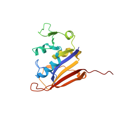
    4ELB, 4ELE, 4ELF, 4ELG, 4ELH

  • PubMed Abstract: 

    Bacterial resistance to antibiotic therapies is increasing and new treatment options are badly needed. There is an overlap between these resistant bacteria and organisms classified as likely bioterror weapons. For example, Bacillus anthracis is innately resistant to the anti-folate trimethoprim due to sequence changes found in the dihydrofolate reductase enzyme. Development of new inhibitors provides an opportunity to enhance the current arsenal of anti-folate antibiotics while also expanding the coverage of the anti-folate class. We have characterized inhibitors of B. anthracis dihydrofolate reductase by measuring the K(i) and MIC values and calculating the energetics of binding. This series contains a core diaminopyrimidine ring, a central dimethoxybenzyl ring, and a dihydrophthalazine moiety. We have altered the chemical groups extended from a chiral center on the dihydropyridazine ring of the phthalazine moiety. The interactions for the most potent compounds were visualized by X-ray structure determination. We find that the potency of individual enantiomers is divergent with clear preference for the S-enantiomer, while maintaining a high conservation of contacts within the binding site. The preference for enantiomers seems to be predicated largely by differential interactions with protein residues Leu29, Gln30 and Arg53. These studies have clarified the activity of modifications and of individual enantiomers, and highlighted the role of the less-active R-enantiomer in effectively diluting the more active S-enantiomer in racemic solutions. This directly contributes to the development of new antimicrobials, combating trimethoprim resistance, and treatment options for potential bioterrorism agents.


  • Organizational Affiliation

    Department of Veterinary Pathobiology, Center for Veterinary Health Sciences, Oklahoma State University, Stillwater, OK 74078, USA. christina.bourne@okstate.edu


Macromolecules
Find similar proteins by:  (by identity cutoff)  |  3D Structure
Entity ID: 1
MoleculeChains Sequence LengthOrganismDetailsImage
Dihydrofolate reductase166Bacillus anthracis str. SterneMutation(s): 0 
Gene Names: BAS2083BA_2237dfrADHFRGBAA_2237
EC: 1.5.1.3
UniProt
Find proteins for Q81R22 (Bacillus anthracis)
Explore Q81R22 
Go to UniProtKB:  Q81R22
Entity Groups  
Sequence Clusters30% Identity50% Identity70% Identity90% Identity95% Identity100% Identity
UniProt GroupQ81R22
Sequence Annotations
Expand
  • Reference Sequence
Small Molecules
Ligands 4 Unique
IDChains Name / Formula / InChI Key2D Diagram3D Interactions
34R
Query on 34R

Download Ideal Coordinates CCD File 
AA [auth F],
DA [auth D],
HA [auth E],
Q [auth C]
(2E)-3-{5-[(2,4-diaminopyrimidin-5-yl)methyl]-2,3-dimethoxyphenyl}-1-[(1R)-1-phenylphthalazin-2(1H)-yl]prop-2-en-1-one
C30 H28 N6 O3
NFQBNKYRVDDBSW-DBQZHCBRSA-N
34S
Query on 34S

Download Ideal Coordinates CCD File 
GA [auth E]
K [auth A]
M [auth H]
T [auth B]
W [auth G]
GA [auth E],
K [auth A],
M [auth H],
T [auth B],
W [auth G],
Z [auth F]
(2E)-3-{5-[(2,4-diaminopyrimidin-5-yl)methyl]-2,3-dimethoxyphenyl}-1-[(1S)-1-phenylphthalazin-2(1H)-yl]prop-2-en-1-one
C30 H28 N6 O3
NFQBNKYRVDDBSW-QSRYBZNWSA-N
CA
Query on CA

Download Ideal Coordinates CCD File 
BA [auth D]
EA [auth E]
I [auth A]
L [auth H]
O [auth C]
BA [auth D],
EA [auth E],
I [auth A],
L [auth H],
O [auth C],
R [auth B],
U [auth G],
X [auth F]
CALCIUM ION
Ca
BHPQYMZQTOCNFJ-UHFFFAOYSA-N
CL
Query on CL

Download Ideal Coordinates CCD File 
CA [auth D]
FA [auth E]
J [auth A]
N [auth H]
P [auth C]
CA [auth D],
FA [auth E],
J [auth A],
N [auth H],
P [auth C],
S [auth B],
V [auth G],
Y [auth F]
CHLORIDE ION
Cl
VEXZGXHMUGYJMC-UHFFFAOYSA-M
Experimental Data & Validation

Experimental Data

  • Method: X-RAY DIFFRACTION
  • Resolution: 2.60 Å
  • R-Value Free: 0.309 
  • R-Value Work: 0.243 
  • R-Value Observed: 0.247 
  • Space Group: P 21 21 21
Unit Cell:
Length ( Å )Angle ( ˚ )
a = 67.874α = 90
b = 135.444β = 90
c = 168.194γ = 90
Software Package:
Software NamePurpose
PHENIXrefinement

Structure Validation

View Full Validation Report


Ligand Structure Quality Assessment 


Entry History 

Deposition Data

Revision History  (Full details and data files)

  • Version 1.0: 2013-02-13
    Type: Initial release
  • Version 1.1: 2023-09-13
    Changes: Data collection, Database references, Derived calculations, Refinement description