4ECJ

Crystal structure of glutathione s-transferase prk13972 (target efi-501853) from pseudomonas aeruginosa pacs2 complexed with glutathione


Experimental Data Snapshot

  • Method: X-RAY DIFFRACTION
  • Resolution: 1.76 Å
  • R-Value Free: 0.183 
  • R-Value Work: 0.163 
  • R-Value Observed: 0.164 

wwPDB Validation   3D Report Full Report


Ligand Structure Quality Assessment 


This is version 1.1 of the entry. See complete history


Literature

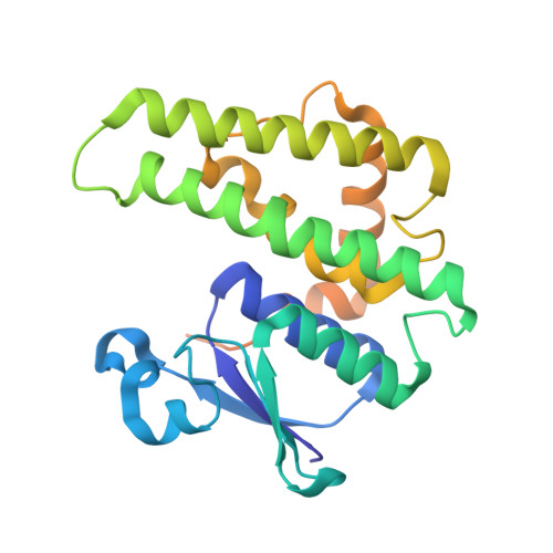
Crystal Structure of Glutathione S-Transferase Prk13972 from Pseudomonas Aeruginosa

Patskovsky, Y.Toro, R.Bhosle, R.Zencheck, W.D.Hillerich, B.Seidel, R.D.Washington, E.Scott Glenn, A.Chowdhury, S.Evans, B.Hammonds, J.Imker, H.J.Armstrong, R.N.Gerlt, J.A.Almo, S.C.

To be published.

Macromolecules
Find similar proteins by:  (by identity cutoff)  |  3D Structure
Entity ID: 1
MoleculeChains Sequence LengthOrganismDetailsImage
glutathione S-transferase
A, B
244Pseudomonas aeruginosa PACS2Mutation(s): 0 
UniProt
Find proteins for A0A0H2Z874 (Pseudomonas aeruginosa (strain UCBPP-PA14))
Explore A0A0H2Z874 
Go to UniProtKB:  A0A0H2Z874
Entity Groups  
Sequence Clusters30% Identity50% Identity70% Identity90% Identity95% Identity100% Identity
UniProt GroupA0A0H2Z874
Sequence Annotations
Expand
  • Reference Sequence
Experimental Data & Validation

Experimental Data

  • Method: X-RAY DIFFRACTION
  • Resolution: 1.76 Å
  • R-Value Free: 0.183 
  • R-Value Work: 0.163 
  • R-Value Observed: 0.164 
  • Space Group: P 21 21 2
Unit Cell:
Length ( Å )Angle ( ˚ )
a = 85.541α = 90
b = 99.857β = 90
c = 51.571γ = 90
Software Package:
Software NamePurpose
PHASERphasing
REFMACrefinement
HKL-3000data reduction
HKL-3000data scaling

Structure Validation

View Full Validation Report


Ligand Structure Quality Assessment 


Entry History 

Revision History  (Full details and data files)

  • Version 1.0: 2012-04-18
    Type: Initial release
  • Version 1.1: 2023-09-13
    Changes: Data collection, Database references, Derived calculations, Refinement description