4EC4

XIAP-BIR3 in complex with a potent divalent Smac mimetic


Experimental Data Snapshot

  • Method: X-RAY DIFFRACTION
  • Resolution: 3.30 Å
  • R-Value Free: 0.276 
  • R-Value Work: 0.210 
  • R-Value Observed: 0.213 

wwPDB Validation   3D Report Full Report


Ligand Structure Quality Assessment 


This is version 1.1 of the entry. See complete history


Literature

Structural insight into inhibitor of apoptosis proteins recognition by a potent divalent smac-mimetic.

Cossu, F.Milani, M.Vachette, P.Malvezzi, F.Grassi, S.Lecis, D.Delia, D.Drago, C.Seneci, P.Bolognesi, M.Mastrangelo, E.

(2012) PLoS One 7: e49527-e49527

  • DOI: https://doi.org/10.1371/journal.pone.0049527
  • Primary Citation of Related Structures:  
    4EB9, 4EC4

  • PubMed Abstract: 

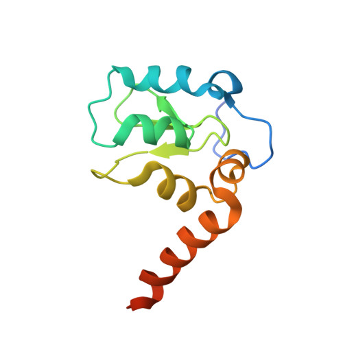
    Genetic alterations enhancing cell survival and suppressing apoptosis are hallmarks of cancer that significantly reduce the efficacy of chemotherapy or radiotherapy. The Inhibitor of Apoptosis Protein (IAP) family hosts conserved proteins in the apoptotic pathway whose over-expression, frequently found in tumours, potentiates survival and resistance to anticancer agents. In humans, IAPs comprise eight members hosting one or more structural Baculoviral IAP Repeat (BIR) domains. Cellular IAPs (cIAP1 and 2) indirectly inhibit caspase-8 activation, and regulate both the canonical and the non-canonical NF-κB signaling pathways. In contrast to cIAPs, XIAP (X chromosome-linked Inhibitor of Apoptosis Protein) inhibits directly the effector caspases-3 and -7 through its BIR2 domain, and initiator caspase-9 through its BIR3 domain; molecular docking studies suggested that Smac/DIABLO antagonizes XIAP by simultaneously targeting both BIR2 and BIR3 domains. Here we report analytical gel filtration, crystallographic and SAXS experiments on cIAP1-BIR3, XIAP-BIR3 and XIAP-BIR2BIR3 domains, alone and in the presence of compound 9a, a divalent homodimeric Smac mimetic. 9a is shown to bind two BIR domains inter- (in the case of two BIR3) and intra-molecularly (in the case of XIAP-BIR2BIR3), with higher affinity for cIAP1-BIR3, relative to XIAP-BIR3. Despite the different crystal lattice packing, 9a maintains a right handed helical conformation in both cIAP1-BIR3 and XIAP-BIR3 crystals, that is likely conserved in solution as shown by SAXS data. Our structural results demonstrate that the 9a linker length, its conformational degrees of freedom and its hydrophobicity, warrant an overall compact structure with optimal solvent exposure of its two active moieties for IAPs binding. Our results show that 9a is a good candidate for pre-clinical and clinical studies, worth of further investigations in the field of cancer therapy.


  • Organizational Affiliation

    Department of Biosciences, University of Milano, Milano, Italy.


Macromolecules
Find similar proteins by:  (by identity cutoff)  |  3D Structure
Entity ID: 1
MoleculeChains Sequence LengthOrganismDetailsImage
Baculoviral IAP repeat-containing protein 4122Homo sapiensMutation(s): 0 
Gene Names: XIAPAPI3BIRC4IAP3
EC: 6.3.2
UniProt & NIH Common Fund Data Resources
Find proteins for P98170 (Homo sapiens)
Explore P98170 
Go to UniProtKB:  P98170
PHAROS:  P98170
GTEx:  ENSG00000101966 
Entity Groups  
Sequence Clusters30% Identity50% Identity70% Identity90% Identity95% Identity100% Identity
UniProt GroupP98170
Sequence Annotations
Expand
  • Reference Sequence
Small Molecules
Ligands 4 Unique
IDChains Name / Formula / InChI Key2D Diagram3D Interactions
0O6
Query on 0O6

Download Ideal Coordinates CCD File 
L [auth A],
O [auth B],
R [auth D],
U [auth E],
W [auth J]
(3S,6S,7S,9aS,3'S,6'S,7'S,9a'S)-N,N'-(benzene-1,4-diylbis{butane-4,1-diyl-1H-1,2,3-triazole-1,4-diyl[(S)-phenylmethanediyl]})bis[7-(hydroxymethyl)-6-{[(2S)-2-(methylamino)butanoyl]amino}-5-oxooctahydro-1H-pyrrolo[1,2-a]azepine-3-carboxamide]
C64 H88 N14 O8
XKEIDWZKJDOBEL-OKDMWEIPSA-N
2PE
Query on 2PE

Download Ideal Coordinates CCD File 
BA [auth G],
V [auth E]
NONAETHYLENE GLYCOL
C18 H38 O10
YZUUTMGDONTGTN-UHFFFAOYSA-N
P33
Query on P33

Download Ideal Coordinates CCD File 
M [auth A],
S [auth D],
Y [auth J]
3,6,9,12,15,18-HEXAOXAICOSANE-1,20-DIOL
C14 H30 O8
XPJRQAIZZQMSCM-UHFFFAOYSA-N
ZN
Query on ZN

Download Ideal Coordinates CCD File 
AA [auth G]
CA [auth K]
DA [auth L]
K [auth A]
N [auth B]
AA [auth G],
CA [auth K],
DA [auth L],
K [auth A],
N [auth B],
P [auth C],
Q [auth D],
T [auth E],
X [auth J],
Z [auth F]
ZINC ION
Zn
PTFCDOFLOPIGGS-UHFFFAOYSA-N
Binding Affinity Annotations 
IDSourceBinding Affinity
0O6 BindingDB:  4EC4 IC50: 25.4 (nM) from 1 assay(s)
PDBBind:  4EC4 IC50: 25.4 (nM) from 1 assay(s)
Experimental Data & Validation

Experimental Data

  • Method: X-RAY DIFFRACTION
  • Resolution: 3.30 Å
  • R-Value Free: 0.276 
  • R-Value Work: 0.210 
  • R-Value Observed: 0.213 
  • Space Group: P 21 21 21
Unit Cell:
Length ( Å )Angle ( ˚ )
a = 65.42α = 90
b = 130.49β = 90
c = 215.73γ = 90
Software Package:
Software NamePurpose
ADSCdata collection
MOLREPphasing
REFMACrefinement
MOSFLMdata reduction
SCALAdata scaling

Structure Validation

View Full Validation Report


Ligand Structure Quality Assessment 


Entry History 

Deposition Data

Revision History  (Full details and data files)

  • Version 1.0: 2012-12-05
    Type: Initial release
  • Version 1.1: 2023-09-13
    Changes: Data collection, Database references, Derived calculations, Refinement description, Structure summary