4E84

Crystal Structure of Burkholderia cenocepacia HldA


Experimental Data Snapshot

  • Method: X-RAY DIFFRACTION
  • Resolution: 2.60 Å
  • R-Value Free: 0.263 
  • R-Value Work: 0.224 
  • R-Value Observed: 0.226 

wwPDB Validation   3D Report Full Report


Ligand Structure Quality Assessment 


This is version 1.3 of the entry. See complete history


Literature

Structural-functional studies of Burkholderia cenocepacia D-glycero-beta-D-manno-heptose 7-phosphate kinase (HldA) and characterization of inhibitors with antibiotic adjuvant and antivirulence properties.

Lee, T.W.Verhey, T.B.Antiperovitch, P.A.Atamanyuk, D.Desroy, N.Oliveira, C.Denis, A.Gerusz, V.Drocourt, E.Loutet, S.A.Hamad, M.A.Stanetty, C.Andres, S.N.Sugiman-Marangos, S.Kosma, P.Valvano, M.A.Moreau, F.Junop, M.S.

(2013) J Med Chem 56: 1405-1417

  • DOI: https://doi.org/10.1021/jm301483h
  • Primary Citation of Related Structures:  
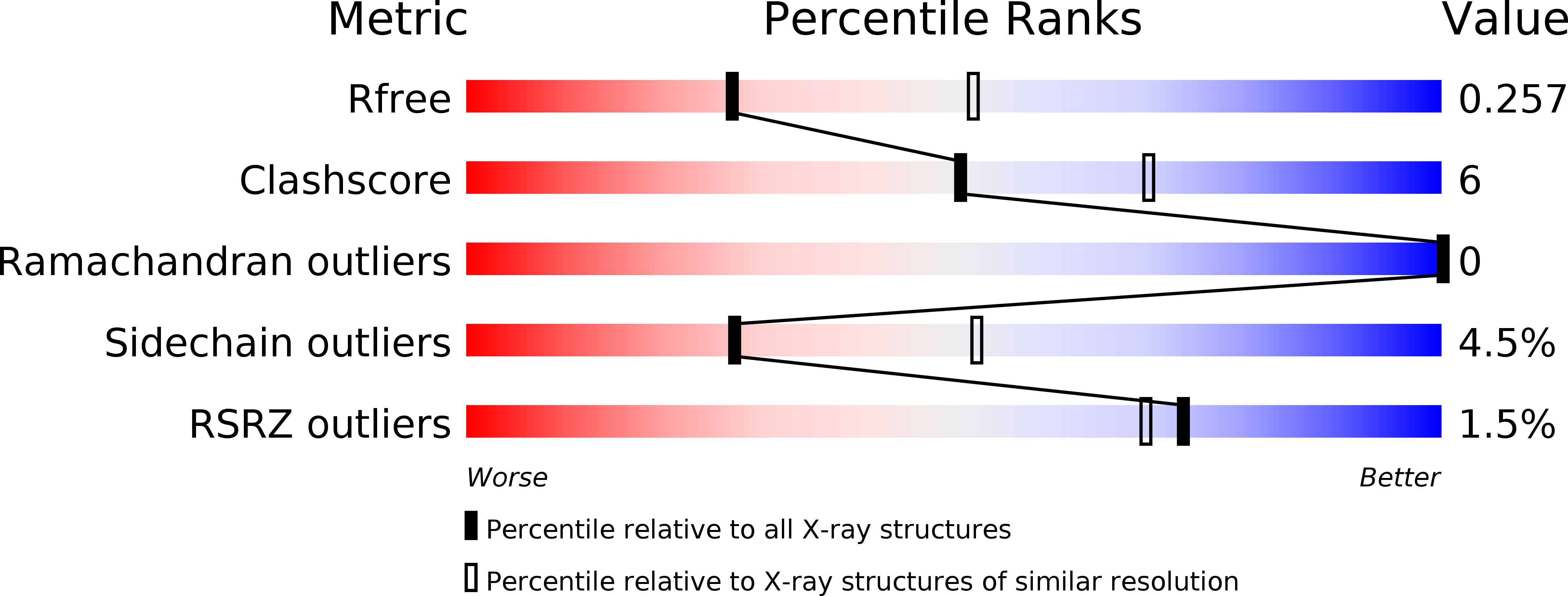
    4E84, 4E8W, 4E8Y, 4E8Z

  • PubMed Abstract: 

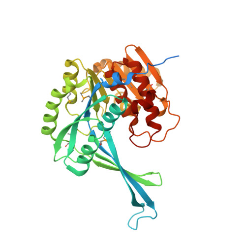
    As an essential constituent of the outer membrane of Gram-negative bacteria, lipopolysaccharide contributes significantly to virulence and antibiotic resistance. The lipopolysaccharide biosynthetic pathway therefore serves as a promising therapeutic target for antivirulence drugs and antibiotic adjuvants. Here we report the structural-functional studies of D-glycero-β-D-manno-heptose 7-phosphate kinase (HldA), an absolutely conserved enzyme in this pathway, from Burkholderia cenocepacia. HldA is structurally similar to members of the PfkB carbohydrate kinase family and appears to catalyze heptose phosphorylation via an in-line mechanism mediated mainly by a conserved aspartate, Asp270. Moreover, we report the structures of HldA in complex with two potent inhibitors in which both inhibitors adopt a folded conformation and occupy the nucleotide-binding sites. Together, these results provide important insight into the mechanism of HldA-catalyzed heptose phosphorylation and necessary information for further development of HldA inhibitors.


  • Organizational Affiliation

    Department of Biochemistry and Biomedical Sciences and Michael G. DeGroote Institute for Infectious Disease Research, McMaster University, 1280 Main Street West, Hamilton, Ontario, L8S 4K1, Canada.


Macromolecules
Find similar proteins by:  (by identity cutoff)  |  3D Structure
Entity ID: 1
MoleculeChains Sequence LengthOrganismDetailsImage
D-beta-D-heptose 7-phosphate kinase
A, B
352Burkholderia cenocepacia J2315Mutation(s): 0 
Gene Names: hldABceJ2315_28810BCAL2945
UniProt
Find proteins for B4EB35 (Burkholderia cenocepacia (strain ATCC BAA-245 / DSM 16553 / LMG 16656 / NCTC 13227 / J2315 / CF5610))
Explore B4EB35 
Go to UniProtKB:  B4EB35
Entity Groups  
Sequence Clusters30% Identity50% Identity70% Identity90% Identity95% Identity100% Identity
UniProt GroupB4EB35
Sequence Annotations
Expand
  • Reference Sequence
Small Molecules
Ligands 7 Unique
IDChains Name / Formula / InChI Key2D Diagram3D Interactions
ANP
Query on ANP

Download Ideal Coordinates CCD File 
C [auth A],
I [auth B]
PHOSPHOAMINOPHOSPHONIC ACID-ADENYLATE ESTER
C10 H17 N6 O12 P3
PVKSNHVPLWYQGJ-KQYNXXCUSA-N
GMZ
Query on GMZ

Download Ideal Coordinates CCD File 
J [auth B]1,7-di-O-phosphono-D-glycero-beta-D-manno-heptopyranose
C7 H16 O13 P2
LMTGTTLGDUACSJ-QTNLNCNHSA-N
M7B
Query on M7B

Download Ideal Coordinates CCD File 
D [auth A]7-O-phosphono-D-glycero-beta-D-manno-heptopyranose
C7 H15 O10 P
SDADNVAZGVDAIM-ZUHYCWGWSA-N
PO4
Query on PO4

Download Ideal Coordinates CCD File 
G [auth A],
M [auth B]
PHOSPHATE ION
O4 P
NBIIXXVUZAFLBC-UHFFFAOYSA-K
K
Query on K

Download Ideal Coordinates CCD File 
E [auth A],
K [auth B]
POTASSIUM ION
K
NPYPAHLBTDXSSS-UHFFFAOYSA-N
CL
Query on CL

Download Ideal Coordinates CCD File 
F [auth A],
L [auth B]
CHLORIDE ION
Cl
VEXZGXHMUGYJMC-UHFFFAOYSA-M
MG
Query on MG

Download Ideal Coordinates CCD File 
H [auth A],
N [auth B]
MAGNESIUM ION
Mg
JLVVSXFLKOJNIY-UHFFFAOYSA-N
Modified Residues  1 Unique
IDChains TypeFormula2D DiagramParent
MSE
Query on MSE
A, B
L-PEPTIDE LINKINGC5 H11 N O2 SeMET
Experimental Data & Validation

Experimental Data

  • Method: X-RAY DIFFRACTION
  • Resolution: 2.60 Å
  • R-Value Free: 0.263 
  • R-Value Work: 0.224 
  • R-Value Observed: 0.226 
  • Space Group: P 61 2 2
Unit Cell:
Length ( Å )Angle ( ˚ )
a = 90.398α = 90
b = 90.398β = 90
c = 397.917γ = 120
Software Package:
Software NamePurpose
CBASSdata collection
PHASERphasing
PHENIXrefinement
HKL-2000data reduction
HKL-2000data scaling

Structure Validation

View Full Validation Report


Ligand Structure Quality Assessment 


Entry History 

Deposition Data

Revision History  (Full details and data files)

  • Version 1.0: 2012-12-26
    Type: Initial release
  • Version 1.1: 2013-06-26
    Changes: Database references
  • Version 1.2: 2013-08-28
    Changes: Database references
  • Version 1.3: 2020-07-29
    Type: Remediation
    Reason: Carbohydrate remediation
    Changes: Data collection, Database references, Derived calculations