4E5P

Thermostable phosphite dehydrogenase A176R variant in complex with NAD


Experimental Data Snapshot

  • Method: X-RAY DIFFRACTION
  • Resolution: 1.90 Å
  • R-Value Free: 0.241 
  • R-Value Work: 0.200 
  • R-Value Observed: 0.202 

wwPDB Validation   3D Report Full Report


Ligand Structure Quality Assessment 


This is version 1.3 of the entry. See complete history


Literature

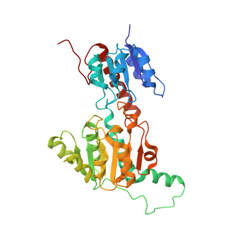
Crystal structures of phosphite dehydrogenase provide insights into nicotinamide cofactor regeneration.

Zou, Y.Zhang, H.Brunzelle, J.S.Johannes, T.W.Woodyer, R.Hung, J.E.Nair, N.van der Donk, W.A.Zhao, H.Nair, S.K.

(2012) Biochemistry 51: 4263-4270

  • DOI: https://doi.org/10.1021/bi2016926
  • Primary Citation of Related Structures:  
    4E5K, 4E5M, 4E5N, 4E5P, 4EBF

  • PubMed Abstract: 

    The enzyme phosphite dehydrogenase (PTDH) catalyzes the NAD(+)-dependent conversion of phosphite to phosphate and represents the first biological catalyst that has been shown to conduct the enzymatic oxidation of phosphorus. Despite investigation for more than a decade into both the mechanism of its unusual reaction and its utility in cofactor regeneration, there has been a lack of any structural data for PTDH. Here we present the cocrystal structure of an engineered thermostable variant of PTDH bound to NAD(+) (1.7 Å resolution), as well as four other cocrystal structures of thermostable PTDH and its variants with different ligands (all between 1.85 and 2.3 Å resolution). These structures provide a molecular framework for understanding prior mutational analysis and point to additional residues, located in the active site, that may contribute to the enzymatic activity of this highly unusual catalyst.


  • Organizational Affiliation

    Department of Biochemistry, University of Illinois at Urbana-Champaign, 600 South Mathews Avenue, Urbana, IL 61801, USA.


Macromolecules
Find similar proteins by:  (by identity cutoff)  |  3D Structure
Entity ID: 1
MoleculeChains Sequence LengthOrganismDetailsImage
Thermostable phosphite dehydrogenase A176R variant
A, B, C, D, E
A, B, C, D, E, F
332Stutzerimonas stutzeriMutation(s): 0 
UniProt
Find proteins for O69054 (Stutzerimonas stutzeri)
Explore O69054 
Go to UniProtKB:  O69054
Entity Groups  
Sequence Clusters30% Identity50% Identity70% Identity90% Identity95% Identity100% Identity
UniProt GroupO69054
Sequence Annotations
Expand
  • Reference Sequence
Experimental Data & Validation

Experimental Data

  • Method: X-RAY DIFFRACTION
  • Resolution: 1.90 Å
  • R-Value Free: 0.241 
  • R-Value Work: 0.200 
  • R-Value Observed: 0.202 
  • Space Group: P 1 21 1
Unit Cell:
Length ( Å )Angle ( ˚ )
a = 70.298α = 90
b = 122.263β = 96.43
c = 134.322γ = 90
Software Package:
Software NamePurpose
MAR345dtbdata collection
PHASERphasing
REFMACrefinement
HKL-2000data reduction
HKL-2000data scaling

Structure Validation

View Full Validation Report


Ligand Structure Quality Assessment 


Entry History 

Deposition Data

Revision History  (Full details and data files)

  • Version 1.0: 2012-05-30
    Type: Initial release
  • Version 1.1: 2012-06-13
    Changes: Database references
  • Version 1.2: 2024-02-28
    Changes: Data collection, Database references, Derived calculations
  • Version 1.3: 2024-04-03
    Changes: Refinement description