4DJS

Structure of beta-catenin in complex with a stapled peptide inhibitor


Experimental Data Snapshot

  • Method: X-RAY DIFFRACTION
  • Resolution: 3.03 Å
  • R-Value Free: 0.291 
  • R-Value Work: 0.263 
  • R-Value Observed: 0.266 

wwPDB Validation   3D Report Full Report


This is version 1.2 of the entry. See complete history


Literature

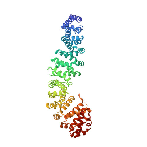
Inhibition of oncogenic Wnt signaling through direct targeting of beta-catenin.

Grossmann, T.N.Yeh, J.T.Bowman, B.R.Chu, Q.Moellering, R.E.Verdine, G.L.

(2012) Proc Natl Acad Sci U S A 109: 17942-17947

  • DOI: https://doi.org/10.1073/pnas.1208396109
  • Primary Citation of Related Structures:  
    4DJS

  • PubMed Abstract: 

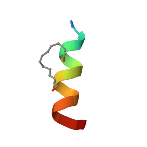
    Aberrant activation of signaling by the Wnt pathway is strongly implicated in the onset and progression of numerous types of cancer. Owing to the persistent dependence of these tumors on Wnt signaling for growth and survival, inhibition of this pathway is considered an attractive mechanism-based therapeutic approach. Oncogenic activation of Wnt signaling can ensue from a variety of distinct aberrations in the signaling pathway, but most share the common feature of causing increased cellular levels of β-catenin by interfering with its constitutive degradation. β-Catenin serves as a central hub in Wnt signaling by engaging in crucial protein-protein interactions with both negative and positive effectors of the pathway. Direct interference with these protein-protein interactions is a biologically compelling approach toward suppression of β-catenin hyperactivity, but such interactions have proven intransigent with respect to small-molecule targeting. Hence β-catenin remains an elusive target for translational cancer therapy. Here we report the discovery of a hydrocarbon-stapled peptide that directly targets β-catenin and interferes with its ability to serve as a transcriptional coactivator for T-cell factor (TCF) proteins, the downstream transcriptional regulators of the Wnt pathway.


  • Organizational Affiliation

    Department of Stem Cell and Regenerative Biology, Harvard University, Cambridge, MA 02138, USA.


Macromolecules
Find similar proteins by:  (by identity cutoff)  |  3D Structure
Entity ID: 1
MoleculeChains Sequence LengthOrganismDetailsImage
Catenin beta-1518Homo sapiensMutation(s): 0 
Gene Names: CTNNB1CTNNBOK/SW-cl.35PRO2286
UniProt & NIH Common Fund Data Resources
Find proteins for P35222 (Homo sapiens)
Explore P35222 
Go to UniProtKB:  P35222
PHAROS:  P35222
GTEx:  ENSG00000168036 
Entity Groups  
Sequence Clusters30% Identity50% Identity70% Identity90% Identity95% Identity100% Identity
UniProt GroupP35222
Sequence Annotations
Expand
  • Reference Sequence

Find similar proteins by:  Sequence   |   3D Structure  

Entity ID: 2
MoleculeChains Sequence LengthOrganismDetailsImage
stapled peptide RRWPQ(MK8)ILD(MK8)HVRRVWR17N/AMutation(s): 0 
Entity Groups  
Sequence Clusters30% Identity50% Identity70% Identity90% Identity95% Identity100% Identity
Sequence Annotations
Expand
  • Reference Sequence
Small Molecules
Modified Residues  1 Unique
IDChains TypeFormula2D DiagramParent
MK8
Query on MK8
B
L-PEPTIDE LINKINGC7 H15 N O2LEU
Experimental Data & Validation

Experimental Data

  • Method: X-RAY DIFFRACTION
  • Resolution: 3.03 Å
  • R-Value Free: 0.291 
  • R-Value Work: 0.263 
  • R-Value Observed: 0.266 
  • Space Group: P 21 21 21
Unit Cell:
Length ( Å )Angle ( ˚ )
a = 63.06α = 90
b = 74.808β = 90
c = 135.714γ = 90
Software Package:
Software NamePurpose
PHENIXrefinement
CNSrefinement
ADSCdata collection
HKL-2000data reduction
SCALEPACKdata scaling
CNSphasing

Structure Validation

View Full Validation Report


Entry History 

Deposition Data

Revision History  (Full details and data files)

  • Version 1.0: 2012-10-17
    Type: Initial release
  • Version 1.1: 2013-08-28
    Changes: Database references
  • Version 1.2: 2019-07-17
    Changes: Data collection, Derived calculations, Refinement description