Structural insights into the specific anti-HIV property of actinohivin: structure of its complex with the alpha(1–2)mannobiose moiety of gp120
Hoque, M.M., Suzuki, K., Tsunoda, M., Jiang, J., Zhang, F., Takahashi, A., Ohbayashi, N., Zhang, X., Tanaka, H., Omura, S., Takenaka, A.(2012) Acta Crystallogr D Biol Crystallogr 68: 1671-1679
- PubMed: 23151632
- DOI: https://doi.org/10.1107/S0907444912040498
- Primary Citation of Related Structures:
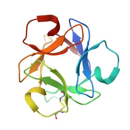
4DEN - PubMed Abstract:
Actinohivin (AH) is an actinomycete lectin with a potent specific anti-HIV activity. In order to clarify the structural evidence for its specific binding to the α(1-2)mannobiose (MB) moiety of the D1 chains of high-mannose-type glycans (HMTGs) attached to HIV-1 gp120, the crystal structure of AH in complex with MB has been determined. The AH molecule is composed of three identical structural modules, each of which has a pocket in which an MB molecule is bound adopting a bracket-shaped conformation. This conformation is stabilized through two weak C-H...O hydrogen bonds facilitated by the α(1-2) linkage. The binding features in the three pockets are quite similar to each other, in accordance with the molecular pseudo-threefold symmetry generated from the three tandem repeats in the amino-acid sequence. The shape of the pocket can accept two neighbouring hydroxyl groups of the O(3) and O(4) atoms of the equatorial configuration of the second mannose residue. To recognize these atoms through hydrogen bonds, an Asp residue is located at the bottom of each pocket. Tyr and Leu residues seem to block the movement of the MB molecules. Furthermore, the O(1) atom of the axial configuration of the second mannose residue protrudes from each pocket into an open space surrounded by the conserved hydrophobic residues, suggesting an additional binding site for the third mannose residue of the branched D1 chain of HMTGs. These structural features provide strong evidence indicating that AH is only highly specific for MB and would facilitate the highly specific affinity of AH for any glycoprotein carrying many HMTGs, such as HIV-1 gp120.
Organizational Affiliation:
Faculty of Pharmacy, Iwaki Meisei University, 5-5-1 Chuodai-Iino, Iwaki, Fukushima, Japan.